CTNNB1 Antikörper (N-Term)
 (8 Referenzen)
 (1 Validierung)
-
- Highlights
-
- Kaninchen-beta-Catenin-Antikörper detektiert zuverlässig beta-Catenin-Ziele in der Wnt-Signalgebung mittels CUT&RUN, ICC, IF, WB, ELISA.
- Gut geeignet für die Darstellung von Adherens Junctions in IHC/IF.
- Hochspezifischer beta-Catenin-Nachweis in WB.
- Target Alle CTNNB1 Antikörper anzeigen
- CTNNB1 (Catenin (Cadherin-Associated Protein), beta 1, 88kDa (CTNNB1))
-
Bindungsspezifität
- N-Term
-
Reaktivität
- Human
-
Wirt
- Kaninchen
-
Klonalität
- Polyklonal
-
Konjugat
- Dieser CTNNB1 Antikörper ist unkonjugiert
-
Applikation
- Western Blotting (WB), Immunohistochemistry (IHC), Immunofluorescence (IF), Immunohistochemistry (Paraffin-embedded Sections) (IHC (p)), Flow Cytometry (FACS), Immunoprecipitation (IP), Immunocytochemistry (ICC), Immunohistochemistry (Frozen Sections) (IHC (fro)), Chromatin Immunoprecipitation (ChIP), Immunohistochemistry (Whole Mount) (IHC (wm)), Cleavage Under Targets and Release Using Nuclease (CUT&RUN)
- Kreuzreaktivität
- Cat, Hund, Human, Maus, Kaninchen, Ratte, Zebrafisch (Danio rerio)
- Produktmerkmale
-
Rabbit Polyclonal antibody to beta Catenin (catenin (cadherin-associated protein), beta 1, 88 kDa)
beta Catenin antibody [N1N2-2], N-term - Aufreinigung
- Purified by antigen-affinity chromatography.
- Güteklasse
- KO Validated
- Immunogen
- Recombinant protein encompassing a sequence within the N-terminus region of human beta Catenin. The exact sequence is proprietary.
- Isotyp
- IgG
- Produktspezifische Information
-
Wofür kann der beta-Catenin-Antikörper ABIN2855042 verwendet werden? Dieser polyklonale, unkonjugierte Kaninchen anti-beta Catenin Antikörper (CATNB, Catenin beta-1 oder β-Catenin) eignet sich für den Nachweis von beta-Catenin (CTNNB1, Catenin beta-1) durch Chromatin-Immunopräzipitation (ChIP), CUT&RUN, Immunfluoreszenz, ELISA, IHC und Western Blotting. Er wurde für die Spezies Mensch, Maus und Ratte validiert. Weitere voraussichtliche Reaktivitäten sind Huhn, Schwein, Xenopus laevis, Rhesusaffe und Zebrafisch.
Welche Validierungsdaten liegen für diesen beta-Catenin-Antikörper vor? Der beta-Catenin-Antikörper verfügt derzeit über eine unabhängige Validierung für CUT&RUN (Cleavage Under Targets and Release Using Nuclease), die im unten stehenden Protokoll nachgelesen werden kann. Er wurde in sieben Publikationen verwendet, die ebenfalls angegeben sind und auf PubMed gefunden werden können. Der Kaninchen-beta-Catenin-Antikörper hat 16 Produktbilder, die seine Leistungsfähigkeit in CUT&RUN, Western Blot, ICC / IF und IHC demonstrieren. Nutzen Sie unseren hochwertigen beta Catenin-Antikörper für den zuverlässigen Nachweis von beta Catenin / β-Catenin beim Menschen und verschiedenen anderen Spezies.
Was ist die Funktion von beta-Catenin? Beta-Catenin ist eine wichtige nachgeschaltete Komponente des kanonischen Wnt-Signalweges. In Abwesenheit von Wnt bildet es einen Komplex mit AXIN1, AXIN2, APC, CSNK1A1 und GSK3B, der die Phosphorylierung an den N-terminalen Ser- und Thr-Resten und die Ubiquitinierung von beta-Catenin über BTRC und dessen anschließenden Abbau durch das Proteasom fördert. In Anwesenheit von Wnt-Liganden wird beta-Catenin nicht ubiquitiniert und reichert sich im Zellkern an, wo es als Koaktivator für Transkriptionsfaktoren der TCF/LEF-Familie fungiert und zur Aktivierung von Wnt-reaktiven Genen führt. (UniProt) Es ist ein schwieriges Ziel für ChIP-seq, aber Sie können unseren beta-Catenin-Antikörper in CUT&RUN verwenden.
-
anti-Catenin (Cadherin-Associated Protein), beta 1, 88kDa (CTNNB1) (AA 692-721), (C-Term) antibody
CTNNB1 Reaktivität: Human WB, IF, IHC (p), FACS Wirt: Kaninchen Polyclonal RB41124 unconjugated
anti-Catenin (Cadherin-Associated Protein), beta 1, 88kDa (CTNNB1) (N-Term) antibodyCTNNB1 Reaktivität: Human, Maus, Ratte WB, IHC, ELISA, IF, ICC Wirt: Kaninchen Polyclonal unconjugated
anti-Catenin (Cadherin-Associated Protein), beta 1, 88kDa (CTNNB1) (AA 2-781) antibodyCTNNB1 Reaktivität: Human WB, IHC, ELISA, IF, IP Wirt: Kaninchen Polyclonal unconjugated
anti-Catenin (Cadherin-Associated Protein), beta 1, 88kDa (CTNNB1) (AA 78-106), (N-Term) antibodyCTNNB1 Reaktivität: Human WB, IF, IHC (p), FACS Wirt: Kaninchen Polyclonal RB41155 unconjugated
anti-Catenin (Cadherin-Associated Protein), beta 1, 88kDa (CTNNB1) (AA 682-781) antibodyCTNNB1 Reaktivität: Human WB, ELISA, IF, PLA, RNAi Wirt: Maus Monoclonal 1C9 unconjugated
anti-Catenin (Cadherin-Associated Protein), beta 1, 88kDa (CTNNB1) (AA 1-100) antibodyCTNNB1 Reaktivität: Human, Maus WB, IHC, ELISA, FACS, ICC, Neut Wirt: Maus Monoclonal 7C5A2 unconjugated
anti-Catenin (Cadherin-Associated Protein), beta 1, 88kDa (CTNNB1) (AA 632-781) antibodyCTNNB1 Reaktivität: Human, Maus, Ratte, Affe WB, IHC, ELISA, FACS Wirt: Maus Monoclonal 5B6E9 unconjugated
anti-Catenin (Cadherin-Associated Protein), beta 1, 88kDa (CTNNB1) antibodyCTNNB1 Reaktivität: Human WB, IHC, IF, FACS, StM Wirt: Kaninchen Monoclonal CTNNB1-2030R unconjugated Recombinant Antibody
anti-Catenin (Cadherin-Associated Protein), beta 1, 88kDa (CTNNB1) antibodyCTNNB1 Reaktivität: Human, Maus, Ratte, Hund, Affe WB, IHC, IF, IHC (p) Wirt: Maus Monoclonal 9C1 unconjugated
anti-Catenin (Cadherin-Associated Protein), beta 1, 88kDa (CTNNB1) antibodyCTNNB1 Reaktivität: Human, Ratte, Hund, Affe WB, IHC, IHC (p), FACS Wirt: Maus Monoclonal 9H2 unconjugated
-
- Applikationshinweise
- WB: 1:500-1:20000. ICC/IF: 1:100-1:1000. IHC-P: 1:100-1:1000. FACS: 1:50-1:200. IP: 1:50-1:100. Optimal dilutions/concentrations should be determined by the researcher. Not tested in other applications.
- Kommentare
-
Positive Control: Mouse brain , PC-12 , HeLa
Validation: Comparison, KO/KD, Orthogonal
- Beschränkungen
- Nur für Forschungszwecke einsetzbar
-
- by
- Cantù Lab, Gene Regulation during Development and Disease, Linköping University
- No.
- #104235
- Datum
- 28.06.2021
- Antigen
- beta Catenin
- Chargennummer
- 42536
- Validierte Anwendung
- Cleavage Under Targets and Release Using Nuclease
- Positivkontrolle
Recombinant anti-H3K27me3 CUT&RUN Positive Control antibody (antibodies-online, ABIN6923144)
- Negativkontrolle
- Monoclonal anti-FLAG (Sigma-Aldrich, F3165)
- Bewertung
Passed. ABIN2855042 allows for beta Catenin-targeted digestion using CUT&RUN on HEK-293 nuclei.
- Primärantikörper
- ABIN2855042
- Sekundärantikörper
- Full Protocol
- Cell harvest
- Harvest 500,000 HEK293T cells per antibody to be used at RT.
- Centrifuge cell solution 3 min at 600 x g at RT.
- Remove the liquid carefully.
- Gently resuspend cells in 1 mL Wash Buffer (20 mM HEPES pH 7.5, 150 mM NaCl, 0.5 mM Spermidine, Roche Complete Protease Inhibitor EDTA-free) by pipetting and transfer cell solution to a 2 mL microcentrifuge tube.
- Centrifuge cell solution 3 min at 600 x g at RT and discard the supernatant.
- Repeat twice for a total of three washes.
- Resuspend cell pellet in 1 mL Wash Buffer by gently pipetting.
- Concanavalin A beads preparation
- Prepare one 1.5 mL microcentrifuge tube.
- Gently resuspend the magnetic Concanavalin A Beads (antibodies-online, ABIN6923139).
- Pipette 40 µL Con A Beads slurry for each sample into the 1.5 mL microcentrifuge tube.
- Place the tube on a magnet stand until the fluid is clear. Remove the liquid carefully.
- Remove the microcentrifuge tube from the magnetic stand.
- Pipette 1 mL Binding Buffer (20 mM HEPES pH 7.5, 10 mM KCl, 1 mM CaCl2, 1 mM MnCl2) into each tube and resuspend ConA beads by gentle pipetting.
- Spin down the liquid from the lid with a quick pulse in a table-top centrifuge.
- Place the tubes on a magnet stand until the fluid is clear. Remove the liquid carefully.
- Remove the microcentrifuge tube from the magnetic stand.
- Repeat twice for a total of three washes.
- Gently resuspend the ConA Beads in a volume of Binding Buffer corresponding to the original volume of bead slurry, i.e. 40 µL per sample.
- Nuclei immobilization – binding to Concanavalin A beads
- Carefully vortex the cell suspension and add 10 µL of the Con A beads in Binding Buffer to the cell suspension for each sample.
- Close tube tightly and rotate for 10 min at RT.
- Primary antibody binding
- oDivide nuclei suspension into separate 2 mL microcentrifuge tubes, one for each antibody (500,000 nuclei per sample).
- Place the microcentrifuge tubes on a magnetic stand until the fluid is clear. Remove the liquid carefully.
- Remove the microcentrifuge tubes from the magnetic stand.
- Place each tube at a low angle on the vortex mixer set to a low speed and add 150 µL Digitonin Wash buffer (wash buffer with 0.025% (wt/vol) Digitonin) supplemented with 2 mM EDTA.
- Gently vortex the microcentrifuge tubes until the beads are resuspended.
- Add 1.5 µL antibody (anti-beta Catenin antibody ABIN2855042, anti-H3K27me3 positive control antibody ABIN6923144, and anti-FLAG tag antibody negative control) to the respective tube, corresponding to a 1:100 dilution.
- Rotate the microcentrifuge tubes ON at 4 °C.
- Spin down the liquid and place the tubes on a magnet stand until the fluid is clear. Remove the liquid carefully.
- Remove the microcentrifuge tubes from the magnetic stand.
- Resuspend with 1 mL Digitonin Wash Buffer and mix by inversion. If clumping occurs, gently remove the clumps with a 1 ml pipette tip.
- Repeat once for a total of two washes.
- pAG-MNase Binding
- Place the tubes on a magnet stand until the fluid is clear. Remove the liquid carefully.
- Remove the microcentrifuge tubes from the magnetic stand.
- o Vortex the sample at low speed and add 2.5 µL (0.05 volumes) CUTANA pAG-MNase for ChIC/CUT&RUN Assays (ABIN6950951) in 50 µL Digitonin Wash Buffer per sample, gently resuspending the beads by pipetting.
- Rotate the microcentrifuge tubes for 1 h at 4 °C.
- Spin down the liquid and place the tubes on a magnet stand until the fluid is clear. Remove the liquid carefully.
- Remove the microcentrifuge tubes from the magnetic stand.
- Resuspend with 1 mL Digitonin Wash Buffer and mix by inversion. If clumping occurs, gently remove the clumps with a 1 mL pipette tip.
- Repeat once for a total of two washes.
- MNase digestion and release of pAG-MNase-antibody-chromatin complexes
- Spin down the liquid from the lid with a quick pulse in a table-top centrifuge.
- Place the tubes on a magnet stand until the fluid is clear. Remove the liquid carefully.
- Place each tube at a low angle on the vortex mixer set to a low speed and add 100 μL Digitonin Wash buffer per sample along the side of the tube.
- Place tubes in a heat block, kept on ice, and allow to chill.
- Add 2 μL 0.1 M CaCl2 to each sample.
- Incubate tubes at 0 °C for 30 min.
- Add 100 μL 2xSTOP buffer (340 mM NaCl, 20 mM EDTA, 4 mM EGTA, 0.05% (wt/vol) Digitonin, 100 μg/mL RNAse A, 50 μg/mL Glycogen).
- Incubate tubes at 37 °C for 30 min.
- Place the tubes on a magnet stand until the fluid is clear.
- Transfer the supernatant containing the pA-MNase-bound digested chromatin fragments to fresh 1.5 mL microcentrifuge tubes.
- DNA extraction
- Add 2 µL 10% SDS to a final concentration of 0.1% and 2.5 µL Proteinase K (20 mg/mL) to each supernatant.
- Gently vortex tubes at a low speed of approximately 1,100 rpm.
- Incubate tubes at 50 °C for 1 h.
- Add 200 µL PCI to tube.
- Vortex tubes thoroughly at high speed until the liquid appears milky.
- Centrifuge tubes in a tabletop centrifuge at 16,000 x g at RT for 5 min.
- Carefully transfer to upper aqueous phase to a fresh 1.5 mL microcentrifuge tube containing 2 µL glycogen (diluted 1:10 to 2 mg/mL from the 20 mg/mL stock solution).
- Add 20 µL 3 M NaOAc pH 5.2.
- Add 400 µL 100% ethanol.
- Place tubes for at -20 °C ON.
- Centrifuge tubes in a tabletop centrifuge at 16,000 x g at 4 °C for 5min.
- Remove the liquid carefully with a pipette.
- Wash pellet with 1ml 70% ethanol.
- Centrifuge tubes in a tabletop centrifuge at 16,000 x g at 4 °C for 1 min.
- Remove the liquid carefully with a pipette.
- Air-dry the pellet, then dissolve in 30 µL 1 mM Tris-HCl, 0.1 mM EDTA.
- Library preparation and sequencing
- Prepare libraries using KAPA HyperPrep Kit using KAPA Dual-Indexed adapters according to protocol.
- Sequence libraries on an Illumina NextSeq 500 sequencer, using a NextSeq 500/550 High Output Kit v2.5 (75 Cycles), 36bp PE.
- Peak calling
- Map reads to the GRCm38 (mm10) mouse genome using Bowtie2 with options: --local --very-sensitive- local --no-unal --no-mixed --no-discordant.
- Call peaks using MACS2 with options -f BAMPE --keep-dup all –nomodel.
- Anmerkungen
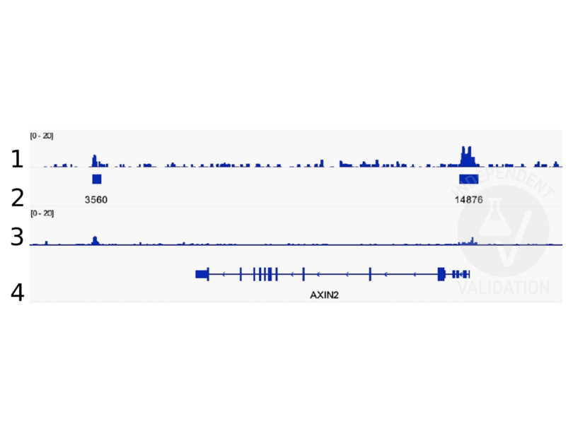
Peaks generated using ABIN2855074 for CUT&RUN aligned well with beta Catenin ChIP-seq tracks(Doumpas et al., 2019).
Validierung #104235 (Cleavage Under Targets and Release Using Nuclease)
 Validierungsbilder
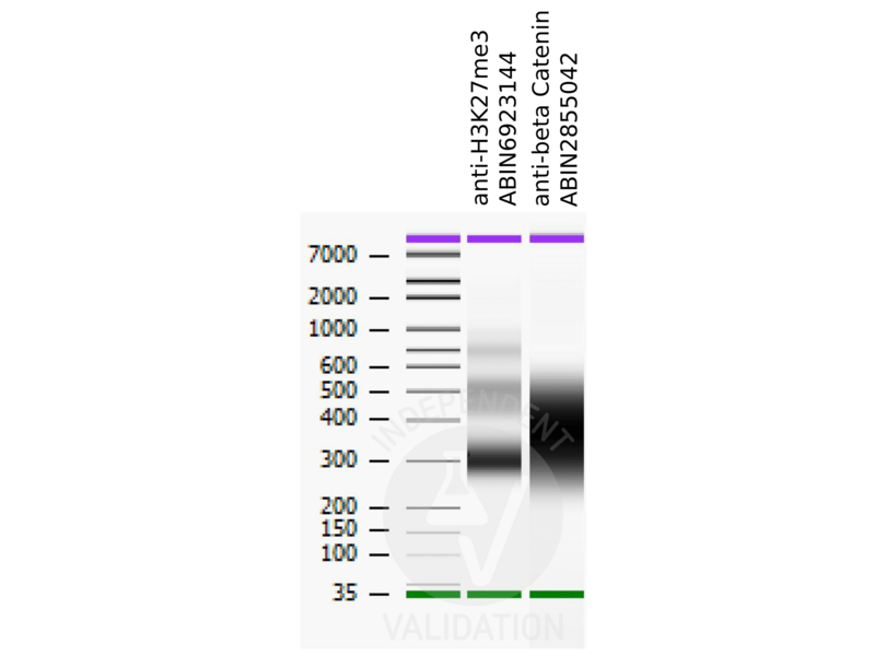
![Bioanalyzer profiles comparing fragment size distributions between reads obtained from CUT&RUN using an anti-H3K27me3 CUT&RUN Positive Control antibody ABIN6923144 and anti-beta Catenin ABIN2855042 after library preparation.]() Bioanalyzer profiles comparing fragment size distributions between reads obtained from CUT&RUN using an anti-H3K27me3 CUT&RUN Positive Control antibody ABIN6923144 and anti-beta Catenin ABIN2855042 after library preparation.
Bioanalyzer profiles comparing fragment size distributions between reads obtained from CUT&RUN using an anti-H3K27me3 CUT&RUN Positive Control antibody ABIN6923144 and anti-beta Catenin ABIN2855042 after library preparation.
![Alignment tracks from CUT&RUN targeting beta Catenin in HEK293T cells. 1. Alignment track for CUT&RUN reads obtained using anti-beta Catenin antibody ABIN2855042 in HEK293T cells. 2. Peaks called by SEACR from CUT&RUN data using anti-beta Catenin antibody ABIN2855042. 3. Peak track from ChIP-seq targeting beta Catenin in HEK293T cells (Doumpas et al., 2019). 4. Refseq track showing the AXIN2 gene on (chr17:65528563-65561648).]() Alignment tracks from CUT&RUN targeting beta Catenin in HEK293T cells. 1. Alignment track for CUT&RUN reads obtained using anti-beta Catenin antibody ABIN2855042 in HEK293T cells. 2. Peaks called by SEACR from CUT&RUN data using anti-beta Catenin antibody ABIN2855042. 3. Peak track from ChIP-seq targeting beta Catenin in HEK293T cells (Doumpas et al., 2019). 4. Refseq track showing the AXIN2 gene on (chr17:65528563-65561648).
Protokoll
Alignment tracks from CUT&RUN targeting beta Catenin in HEK293T cells. 1. Alignment track for CUT&RUN reads obtained using anti-beta Catenin antibody ABIN2855042 in HEK293T cells. 2. Peaks called by SEACR from CUT&RUN data using anti-beta Catenin antibody ABIN2855042. 3. Peak track from ChIP-seq targeting beta Catenin in HEK293T cells (Doumpas et al., 2019). 4. Refseq track showing the AXIN2 gene on (chr17:65528563-65561648).
Protokoll -
- Format
- Liquid
- Konzentration
- 0.07 mg/mL
- Buffer
- 1XPBS ( pH 7), 1 % BSA, 20 % Glycerol, 0.025 % ProClin 300
- Konservierungsmittel
- ProClin
- Vorsichtsmaßnahmen
- This product contains ProClin: a POISONOUS AND HAZARDOUS SUBSTANCE which should be handled by trained staff only.
- Lagerung
- 4 °C,-20 °C
- Informationen zur Lagerung
- Store as concentrated solution. Centrifuge briefly prior to opening vial. For short-term storage (1-2 weeks), store at 4°C. For long-term storage, aliquot and store at -20°C or below. Avoid multiple freeze-thaw cycles.
-
-
: "Acute Intoxication With Alcohol Reduces Trauma-Induced Proinflammatory Response and Barrier Breakdown in the Lung via the Wnt/β-Catenin Signaling Pathway." in: Frontiers in immunology, Vol. 13, pp. 866925, (2022) (PubMed).
: "A new cut&run low volume-urea (LoV-U) protocol optimized for transcriptional co-factors uncovers Wnt/b-catenin tissue-specific genomic targets." in: Development (Cambridge, England), (2022) (PubMed).
: "Identification of candidate lncRNAs and circRNAs regulating WNT3/β-catenin signaling in essential hypertension." in: Aging, Vol. 12, Issue 9, pp. 8261-8288, (2020) (PubMed).
: "A novel c-Kit/phospho-prohibitin axis enhances ovarian cancer stemness and chemoresistance via Notch3-PBX1 and β-catenin-ABCG2 signaling." in: Journal of biomedical science, Vol. 27, Issue 1, pp. 42, (2020) (PubMed).
: "Small G protein signalling modulator 2 (SGSM2) is involved in oestrogen receptor-positive breast cancer metastasis through enhancement of migratory cell adhesion via interaction with E-cadherin." in: Cell adhesion & migration, Vol. 13, Issue 1, pp. 120-137, (2019) (PubMed).
: "Targeting the Wnt/β-catenin pathway in human osteosarcoma cells." in: Oncotarget, Vol. 9, Issue 95, pp. 36780-36792, (2018) (PubMed).
: "ST6GALNAC1 plays important roles in enhancing cancer stem phenotypes of colorectal cancer via the Akt pathway." in: Oncotarget, Vol. 8, Issue 68, pp. 112550-112564, (2017) (PubMed).
: "Control of the negative IRES trans-acting factor KHSRP by ubiquitination." in: Nucleic acids research, Vol. 45, Issue 1, pp. 271-287, (2017) (PubMed).
-
: "Acute Intoxication With Alcohol Reduces Trauma-Induced Proinflammatory Response and Barrier Breakdown in the Lung via the Wnt/β-Catenin Signaling Pathway." in: Frontiers in immunology, Vol. 13, pp. 866925, (2022) (PubMed).
-
- Target
- CTNNB1 (Catenin (Cadherin-Associated Protein), beta 1, 88kDa (CTNNB1))
- Andere Bezeichnung
- catenin beta 1 (CTNNB1 Produkte)
- Synonyme
- Jup antikoerper, LOC100217813 antikoerper, CTNNB1 antikoerper, CTNNB antikoerper, Bfc antikoerper, Catnb antikoerper, Mesc antikoerper, ctnnb antikoerper, id:ibd2058 antikoerper, wu:fb73e10 antikoerper, wu:fi81c06 antikoerper, wu:fk25h01 antikoerper, ctnnb1 antikoerper, CHBCAT antikoerper, MRD19 antikoerper, armadillo antikoerper, beta-catenin antikoerper, catenin beta 1 antikoerper, catenin (cadherin associated protein), beta 1 antikoerper, catenin (cadherin-associated protein), beta 1 antikoerper, catenin beta 1 S homeolog antikoerper, catenin beta 1 L homeolog antikoerper, CTNNB1 antikoerper, Ctnnb1 antikoerper, ctnnb1 antikoerper, ctnnb1.S antikoerper, ctnnb1.L antikoerper
- Hintergrund
-
Beta-catenin is an adherens junction protein. Adherens junctions (AJs, also called the zonula adherens) are critical for the establishment and maintenance of epithelial layers, such as those lining organ surfaces. AJs mediate adhesion between cells, communicate a signal that neighboring cells are present, and anchor the actin cytoskeleton. In serving these roles, AJs regulate normal cell growth and behavior. At several stages of embryogenesis, wound healing, and tumor cell metastasis, cells form and leave epithelia. This process, which involves the disruption and reestablishment of epithelial cell-cell contacts, may be regulated by the disassembly and assembly of AJs. AJs may also function in the transmission of the 'contact inhibition' signal, which instructs cells to stop dividing once an epithelial sheet is complete.[supplied by OMIM]
Cellular Localization: Cytoplasm , Nucleus , cytoskeleton - Molekulargewicht
- 85 kDa
- Gen-ID
- 1499
- UniProt
- P35222
- Pathways
- WNT Signalweg, Intracellular Steroid Hormone Receptor Signaling Pathway, Peptide Hormone Metabolism, Regulation of Muscle Cell Differentiation, Cell-Cell Junction Organization, Tube Formation, Maintenance of Protein Location, Signaling Events mediated by VEGFR1 and VEGFR2
-


