TNFSF9 Antikörper (AA 101-200)
 (1 Validierung)
-
- Target Alle TNFSF9 Antikörper anzeigen
- TNFSF9 (Tumor Necrosis Factor (Ligand) Superfamily, Member 9 (TNFSF9))
-
Bindungsspezifität
- AA 101-200
- Reaktivität
- Human, Maus, Ratte
-
Wirt
- Kaninchen
-
Klonalität
- Polyklonal
-
Konjugat
- Dieser TNFSF9 Antikörper ist unkonjugiert
-
Applikation
- Western Blotting (WB), ELISA, Immunocytochemistry (ICC), Immunofluorescence (Paraffin-embedded Sections) (IF (p)), Immunofluorescence (Cultured Cells) (IF (cc)), Immunohistochemistry (Paraffin-embedded Sections) (IHC (p)), Immunohistochemistry (Frozen Sections) (IHC (fro))
- Kreuzreaktivität
- Human, Maus, Ratte
- Aufreinigung
- Purified by Protein A.
- Immunogen
- KLH conjugated synthetic peptide derived from human TNFSF9
- Isotyp
- IgG
-
anti-Tumor Necrosis Factor (Ligand) Superfamily, Member 9 (TNFSF9) (AA 42-253) antibody
TNFSF9 Reaktivität: Human WB, IHC, IP, ICC Wirt: Kaninchen Polyclonal unconjugated
anti-Tumor Necrosis Factor (Ligand) Superfamily, Member 9 (TNFSF9) (AA 105-309) antibodyTNFSF9 Reaktivität: Maus WB, IHC, IP, ICC Wirt: Kaninchen Polyclonal unconjugated
anti-Tumor Necrosis Factor (Ligand) Superfamily, Member 9 (TNFSF9) (AA 52-254) antibodyTNFSF9 Reaktivität: Human ELISA, FACS Wirt: Kaninchen Monoclonal DM68 unconjugated Recombinant Antibody
anti-Tumor Necrosis Factor (Ligand) Superfamily, Member 9 (TNFSF9) antibodyTNFSF9 Reaktivität: Human FACS, IHC, StM Wirt: Maus Monoclonal CD137L-1547 unconjugated
anti-Tumor Necrosis Factor (Ligand) Superfamily, Member 9 (TNFSF9) (Extracellular Domain) antibodyTNFSF9 Reaktivität: Human WB, ELISA, FACS, ICC Wirt: Maus Monoclonal 41B436 unconjugated
anti-Tumor Necrosis Factor (Ligand) Superfamily, Member 9 (TNFSF9) (AA 52-254) antibodyTNFSF9 Reaktivität: Human ELISA, IHC, IF Wirt: Kaninchen Polyclonal unconjugated
anti-Tumor Necrosis Factor (Ligand) Superfamily, Member 9 (TNFSF9) (AA 145-254) antibodyTNFSF9 Reaktivität: Human WB, ELISA Wirt: Maus Monoclonal 1D7 unconjugated
anti-Tumor Necrosis Factor (Ligand) Superfamily, Member 9 (TNFSF9) (AA 145-254) antibodyTNFSF9 Reaktivität: Human WB, ELISA Wirt: Maus Polyclonal unconjugated
anti-Tumor Necrosis Factor (Ligand) Superfamily, Member 9 (TNFSF9) antibodyTNFSF9 Reaktivität: Maus WB, FACS, BP Wirt: Kaninchen Chimeric AT113-2 unconjugated Recombinant Antibody
anti-Tumor Necrosis Factor (Ligand) Superfamily, Member 9 (TNFSF9) antibodyTNFSF9 Reaktivität: Maus FACS, BR Wirt: Kaninchen Chimeric TKS-1 unconjugated Recombinant Antibody
-
- Applikationshinweise
-
WB 1:300-5000
ELISA 1:500-1000
IHC-P 1:200-400
IHC-F 1:100-500
IF(IHC-P) 1:50-200
IF(IHC-F) 1:50-200
IF(ICC) 1:50-200
ICC 1:100-500 - Beschränkungen
- Nur für Forschungszwecke einsetzbar
-
- by
- Immunohistochemistry Core, NYU Langone
- No.
- #029578
- Datum
- 18.01.2014
- Antigen
- Chargennummer
- 999892W
- Validierte Anwendung
- Immunohistochemistry
- Positivkontrolle
- Human liver
- Negativkontrolle
- Human placental decidual cells
- Bewertung
- Signal was detected in positive control tissue and not in negative control tissue.
- Primärantikörper
- Antibody: Tumor necrosis factor ligand superfamily member 9 (TNFSF9)
- Catalog number: ABIN705056
- Lot number: 999892W
- Sekundärantikörper
- Antibody: Biotinylated goat anti-rabbit/anti-mouse (Kit)
- Lot number: D05923BA
- Full Protocol
- Immunohistochemistry was performed on a Ventana NexES automated platform, instrument manufacturer specific reagents are italicized.
- 1. Slides were preheated in convection oven at 60°C for 30 minutes
- 2. Deparaffinization procedure: - 3 changes of Xylene, 5 minutes each - 3 changes of 100% Ethanol, 3 minutes each - 3 changes of 95% Ethanol, 3 minutes each - Rinsed in distilled water, 3 changes
- 1. Heat retrieval procedure - Slides retrieved in 10.0 mM Citrate, pH6.0 in a 1000W microwave oven (~100°C) for 15 minutes. - Slides were allowed to cool (in citrate) for 30 minutes. - Slides were washed x 3 in Distilled water
- 1. NexES instrument procedure, iVIEW DAB paraffin protocol (*abridged*): - Slide chamber warmed to 37°C
- 1. Slides rinsed with *reaction buffer* x 3
- 1. *iVIEW Inhibitor (H2O2)* applied and incubated for 4 minutes
- 1. Slides rinsed with *reaction buffer*
- 1. Antibody Application - Primary antibody diluted 1:250 in PBS (100 microliters applied/slide) - Ventana Isotype control applied neat - Slides incubated overnight at room temperature (~12 hours ~25°C)
- 1. Slides rinsed with *reaction buffer* x3
- 1. *iVIEW Biotinylated IgG* applied and incubated for 8 minutes
- 1. Slides rinsed with *reaction buffer*
- 1. *iVIEW Streptavidin-Horseradish Peroxidase* applied and incubated for 8 minutes
- 1. Slides rinsed with *reaction buffer*
- 1. *iVIEW DAB/H2O2* applied and incubated for 8 minutes
- 1. Slides rinsed with *reaction buffer*
- 1. *iVIEW Copper* applied and incubated for 4 minutes
- 1. Slides rinsed with *reaction buffer*
- 1. Slides washed in Dawn Detergent/tap water
- 1. Counterstain Procedure - Hematoxylin (Leica 560 MX) 30 seconds - Slides washed in tap water, 1 minute - Decolorized (10% Acetic Acid in 70% ethanol), 1 minute - Slides washed in tap water, 1 minute - Bluing (Austin Clear Ammonia), 1 minute - Slides washed in tap water, 1 minute
- 1. Dehydration/coverslipping procedure: - 3 changes of 95% Ethanol, 3 minutes each - 3 changes of 100% Ethanol, 3 minutes each - 3 changes of Xylene, 5 minutes each - Mounted with Permount
- 1. Imaging: Leica SCN 400F Whole Slide Scanner with Digital Image Hub and Leica Slidepath software
- Anmerkungen
- Deviations from protocol/procedure supplied by manufacturer:
- Step 1: Heated tissue 60°C for 30 minutes; manufacturer heats for 45 minutes.
- Step 2: No ethanol wash was performed during deparaffinization; manufacturer includes 1 wash of 80% ethanol for 3 minutes.
- Step 3.1: Slides heated for 15 minutes; manufacturer provides a range of 15-20 minutes.
- Step 3.2: Slides cooled for 30 minutes; manufacturer cools for 20 minutes.
- Step 4: Italicized reagents and incubation time are fixed instrument parameters.
- Step 5: Secondary species-specific serum block not used; manufacturer blocks with 5% normal goat serum for 2 hours.
- Step 8.1: Antibody diluted in PBS at 1:250; manufacture recommends dilution range (1:100-1:500). No primary antibody diluent recommended.
- Step 8.2.1: Primary antibody incubated at room temperature overnight; manufacturer incubates overnight 4°C with agitation.
- Tissue Interpretation (limited): • TNFSF9: Under the staining parameters described above, placenta was weakly cytoplasmic positive (trophoblastic and decidua cells). Liver hepatocytes were weakly stained. Substantial signal detected in limited number of other tissues, including kidney, prostate, and skin.
- • I-NC (Isotype negative control): No signal detected
- • B-NC (Blank negative control): No signal detected
- Signal Localization: - Signal to noise was adequate with predominately cytoplasmic subcellular localization observed. Nuclear staining was rare and no distinct membrane signal was detected.
Validierung #029578 (Immunohistochemistry)
 Validierungsbilder
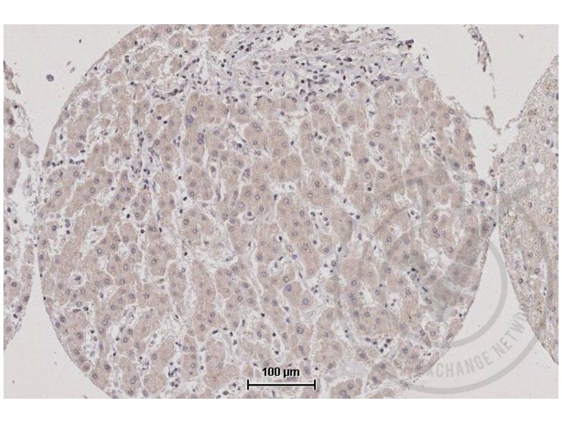
![Figure 1: TNFSF9 staining of human liver (brown). Counterstain in blue.]() Figure 1: TNFSF9 staining of human liver (brown). Counterstain in blue.
Figure 1: TNFSF9 staining of human liver (brown). Counterstain in blue.
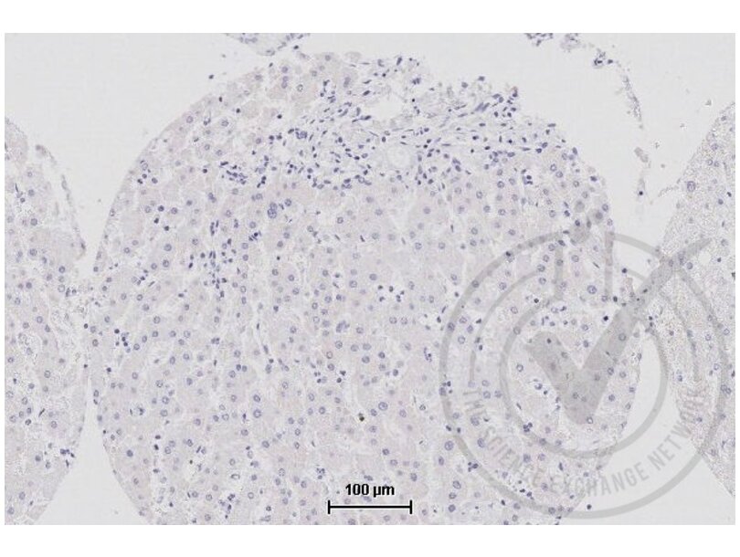
![Figure 2: Isotype control staining of human liver (brown). Counterstain in blue.]() Figure 2: Isotype control staining of human liver (brown). Counterstain in blue.
Figure 2: Isotype control staining of human liver (brown). Counterstain in blue.
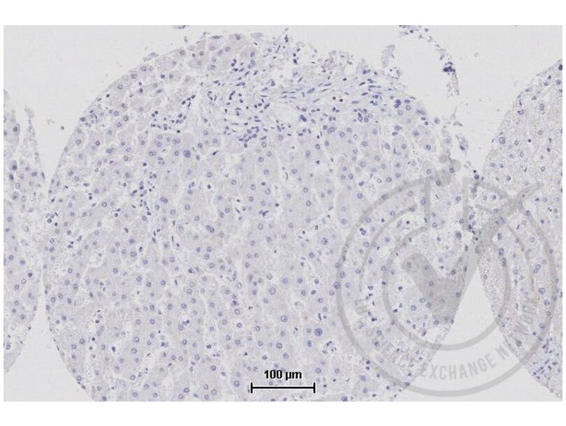
![Figure 3: Secondary only staining of human liver (brown). Counterstain in blue.]() Figure 3: Secondary only staining of human liver (brown). Counterstain in blue.
Figure 3: Secondary only staining of human liver (brown). Counterstain in blue.
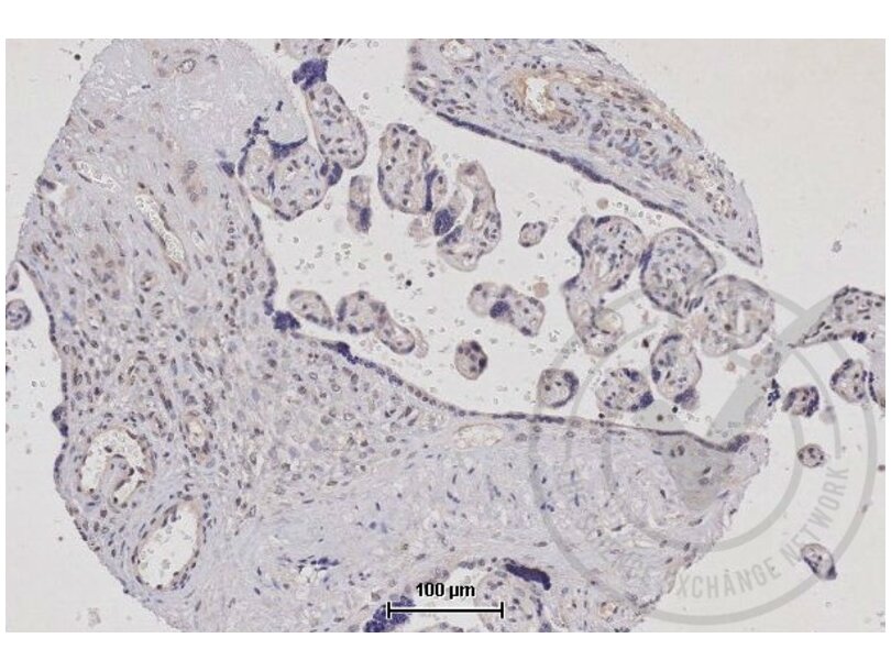
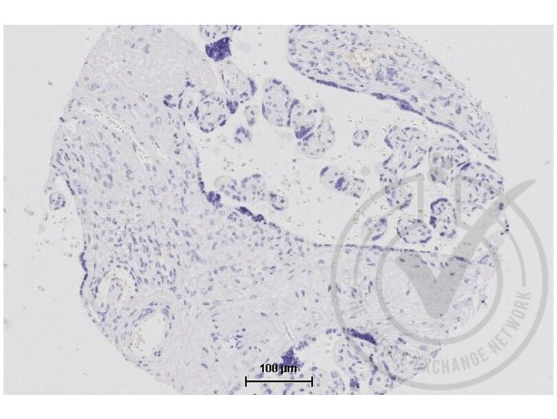
![Figure 4: TNFSF9 staining of human placenta (brown). Counterstain in blue.]() Figure 4: TNFSF9 staining of human placenta (brown). Counterstain in blue.
Figure 4: TNFSF9 staining of human placenta (brown). Counterstain in blue.
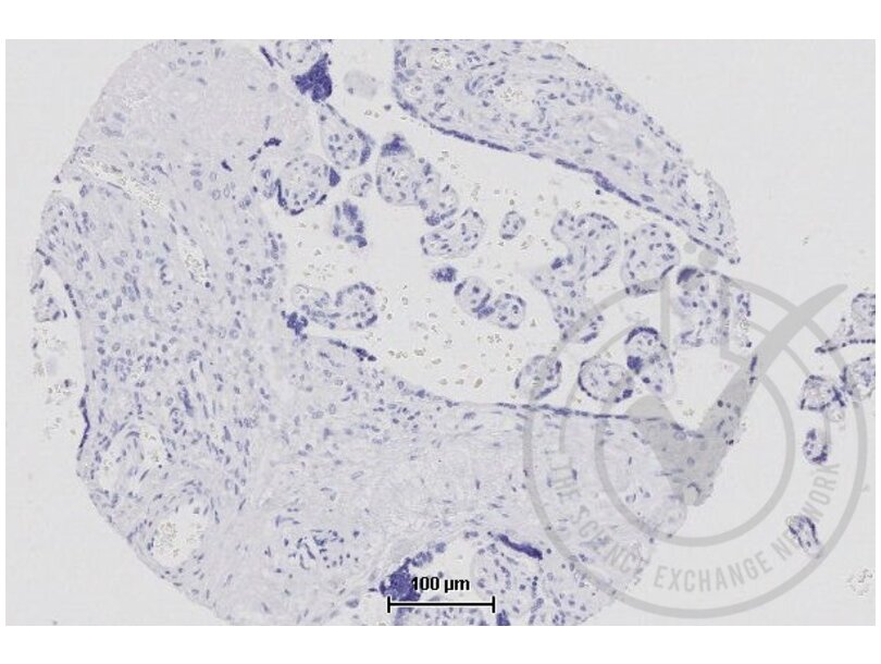
![Figure 5: Isotype control staining of human placenta (brown). Counterstain in blue.]() Figure 5: Isotype control staining of human placenta (brown). Counterstain in blue.
Figure 5: Isotype control staining of human placenta (brown). Counterstain in blue.
![Figure 6: Secondary only staining of human placenta (brown). Counterstain in blue.]() Figure 6: Secondary only staining of human placenta (brown). Counterstain in blue.
Protokoll
Figure 6: Secondary only staining of human placenta (brown). Counterstain in blue.
Protokoll -
- Format
- Liquid
- Konzentration
- 1 μg/μL
- Buffer
- 0.01M TBS( pH 7.4) with 1 % BSA, 0.02 % Proclin300 and 50 % Glycerol.
- Konservierungsmittel
- ProClin
- Vorsichtsmaßnahmen
- This product contains ProClin: a POISONOUS AND HAZARDOUS SUBSTANCE, which should be handled by trained staff only.
- Lagerung
- 4 °C,-20 °C
- Informationen zur Lagerung
- Shipped at 4°C. Store at -20°C for one year. Avoid repeated freeze/thaw cycles.
- Haltbarkeit
- 12 months
-
- Target
- TNFSF9 (Tumor Necrosis Factor (Ligand) Superfamily, Member 9 (TNFSF9))
- Andere Bezeichnung
- TNFSF9/CD137L (TNFSF9 Produkte)
- Synonyme
- TNFSF9 antikoerper, 4-1BB-L antikoerper, 4-1BBL antikoerper, AI848817 antikoerper, Cd137l antikoerper, Ly63l antikoerper, CD137L antikoerper, TNF superfamily member 9 antikoerper, tumor necrosis factor (ligand) superfamily, member 9 antikoerper, Tnfsf9 antikoerper, LOC476729 antikoerper, TNFSF9 antikoerper
- Hintergrund
-
Synonyms: CD137L, 4-1BB-L, Tumor necrosis factor ligand superfamily member 9, 4-1BB ligand, 4-1BBL, TNFSF9
Background: Cytokine that binds to TNFRSF9. Induces the proliferation of activated peripheral blood T-cells. May have a role in activation-induced cell death (AICD). May play a role in cognate interactions between T-cells and B-cells/macrophages.
- Gen-ID
- 8744
- UniProt
- P41273
- Pathways
- Activated T Cell Proliferation, Cancer Immune Checkpoints
-


