RFP Antikörper
 (356 Referenzen)
 (1 Validierung)
-
- Highlights
-
- Häufig zitiert in wissenschaftlichen Veröffentlichungen mit mehr als 300 Zitaten
- Inhouse für relevante Anwendungen validiert
- Ausführlicher Validierungsbericht für IF, erstellt von einem Ihrer Kollegen
- Entwickelt für den Nachweis von RFP und seinen Varianten
- Target Alle RFP Antikörper anzeigen
- RFP (Red Fluorescent Protein (RFP))
- Reaktivität
- Discosoma
-
Wirt
- Kaninchen
-
Klonalität
- Polyklonal
-
Konjugat
- Dieser RFP Antikörper ist unkonjugiert
-
Applikation
- Western Blotting (WB), ELISA, Immunofluorescence (IF), Immunohistochemistry (IHC), Immunoprecipitation (IP), Flow Cytometry (FACS), Immunohistochemistry (Frozen Sections) (IHC (fro)), Immunohistochemistry (Paraffin-embedded Sections) (IHC (p))
- Verwendungszweck
- Polyclonal RFP antibody is designed to detect RFP and its variants.
- Kreuzreaktivität (Details)
-
Expect reactivity against RFP and its variants: mCherry, tdTomato, mBanana, mPlum, mOrange and mStrawberry. Assay by immunoelectrophoresis resulted in a single precipitin arc against anti-Rabbit Serum and purified and partially purified Red Fluorescent Protein (Discosoma).
No reaction was observed against Human, Mouse or Rat serum proteins. - Aufreinigung
- This product was prepared from monospecific antiserum by immunoaffinity chromatography using Red Fluorescent Protein (Discosoma) coupled to agarose beads followed by solid phase adsorption(s) to remove any unwanted reactivities.
- Sterilität
- Sterile filtered
- Immunogen
-
The immunogen is a Red Fluorescent Protein (RFP) fusion protein corresponding to the full length amino acid sequence (234aa) derived from the mushroom polyp coral Discosoma.
Immunogen Type: Recombinant Protein - Isotyp
- IgG
- Produktspezifische Information
-
Wozu kann der RFP-Antikörper ABIN129578 verwendet werden? Dieser polyklonale RFP-Antikörper detektiert RFP und seine Varianten, wie mCherry, tdTomato, mBanana, mOrange und andere. Der RFP-Antikörper wurde für verschiedene Anwendungen validiert und kann für den Nachweis von RFP und Derivaten durch ELISA, Immunfluoreszenz, FACS, Immunhistochemie oder Western Blotting verwendet werden.
Welche Validierungsdaten sind für diesen RFP-Antikörper verfügbar? Der Antikörper wird in mehreren hundert Publikationen referenziert und zeichnet sich durch eine erwiesene, sehr hohe Zuverlässigkeit aus. Er verfügt derzeit über 12 Produktbilder, die seine Leistungsfähigkeit in einer Vielzahl von Anwendungen zeigen. Das Produkt ist derzeit in Mengen von 10µl und 100µl erhältlich. RFP-Antikörper für den Nachweis von rot fluoreszierendem Protein und Derivaten.
Was ist die Funktion von RFP? RFP ist ein Fluorophor, der bei Anregung rot-orange fluoresziert. Das Protein hat viele Anwendungen, unter anderem als Marker bei der Überwachung physiologischer Prozesse in vivo. Das Anregungsmaximum liegt bei etwa 555 nm, das Emissionsmaximum bei 584 nm. RFP-Antikörper bieten eine recht einfach zu handhabende Möglichkeit, RFP in seinen verschiedenen Anwendungen sichtbar zu machen.
-
anti-Red Fluorescent Protein (RFP) antibody
RFP Reaktivität: Discosoma WB, ELISA, IP, FACS, FM Wirt: Maus Monoclonal 8E5-G7 unconjugated
anti-Red Fluorescent Protein (RFP) antibodyRFP Reaktivität: Discosoma WB, IF, IHC (fro), IHC (p) Wirt: Ziege Polyclonal unconjugated
anti-Red Fluorescent Protein (RFP) antibodyRFP Reaktivität: Discosoma WB Wirt: Maus Monoclonal E64-1077 unconjugated
anti-Red Fluorescent Protein (RFP) antibodyRFP Reaktivität: Discosoma WB, ELISA Wirt: Huhn Polyclonal unconjugated
anti-Red Fluorescent Protein (RFP) antibody (DyLight 488)RFP Reaktivität: Discosoma WB, IF, IHC (fro) Wirt: Ziege Polyclonal DyLight 488
anti-Red Fluorescent Protein (RFP) antibody (DyLight 550)RFP Reaktivität: Discosoma WB, IF, IHC (fro) Wirt: Ziege Polyclonal DyLight 550
anti-Red Fluorescent Protein (RFP) antibody (DyLight 633)RFP Reaktivität: Discosoma WB, IF, IHC (fro) Wirt: Ziege Polyclonal DyLight 633
anti-Red Fluorescent Protein (RFP) antibody (Biotin)RFP Reaktivität: Discosoma WB, ELISA, IHC Wirt: Kaninchen Polyclonal Biotin
anti-Red Fluorescent Protein (RFP) antibody (Biotin)RFP Reaktivität: Discosoma WB, ELISA, IF, FM Wirt: Huhn Polyclonal Biotin
anti-Red Fluorescent Protein (RFP) antibody (DyLight 405)RFP Reaktivität: Discosoma WB, IF, IHC (fro) Wirt: Ziege Polyclonal DyLight 405
-
- Applikationshinweise
-
Suggested dilutions:
ELISA: 1:28,700 - 1:48,700
IF: 1:200 - 1:2,000
FACS: 1:200 - 1:2,000
WB: 1:1,000 - 1:5,000
IHC: 1:200 - 1:2,000
IP: User Optimized - Kommentare
-
This RFP antibody can be used to detect RFP by ELISA (sandwich or capture) for the direct binding of antigen.
Optimal titers for applications should be determined by the researcher. - Beschränkungen
- Nur für Forschungszwecke einsetzbar
-
- by
- Centre d'Immunologie de Marseille-Luminy
- No.
- #101103
- Datum
- 07.03.2017
- Antigen
- RFP
- Chargennummer
- 34945
- Validierte Anwendung
- Immunofluorescence
- Positivkontrolle
- CX3CL1mcherry+ murine thymus
- Negativkontrolle
- CX3CL1 WT murine thymus
- Bewertung
Passed. The RFP antibody ABIN129578 specifically labels RFP+ cells in CX3CL1mcherry+ murine thymus in immunofluorescence, consistent with the expression pattern of CX3CL1mcherry.
- Primärantikörper
- ABIN1043867
- Sekundärantikörper
- donkey Fab'2 anti-rabbit A647 conjugated antibody (Jackson Immunoresearch, 711-606-152, lot 128806)
- Full Protocol
- Harvest thymus from 7 weeks old mice in 1X Dulbecco's phosphate buffered saline (DPBS) (Gibco Life Technologies, 14200-067).
- Fix mouse thymus in antigen fix (Diapath, P0014) for 2h at 4?C.
- Wash tissue in 0.1M pH7.4 phosphate buffer at for 1h at 4?C.
- Dehydrate tissue in 30% sucrose solution at 4?C ON.
- Snap freeze tissue in Tissue Freezing Medium (ElectroMicroscopy Science, 72592-C) at -80?C.
- Cut blocks into 25?m sections using a cryostat (Leica, CM3050 S).
- Transfer sections to a slide.
- Create a hydrophobic barrier on the slide around sections with Dako pen (Dako, S2002, lot 00081640).
- Place slide in a humidified chamber and rehydrate sections in 0.1 M TrisHCl pH7.4 for 10min at RT.
- Gently remove buffer by tapping slide.
- Permeabilize tissue in 0.1M TrisHCl pH7.4 containing 2% Triton X-100 (Sigma, lot 015K0039) and 0.5% BSA, for 20min at RT.
- Incubate sections with primary RFP antibody (Red Fluorescent Protein) (AA 234) (antibodies-online, ABIN129578, lot 34945) diluted 1:1000 0.1M TrisHCl pH7.4 containing 2% Triton X-100 (Sigma, batch 015K0039) and 0.5% BSA for 2h at RT.
- Incubate a no primary antibody negative control in parallel in 0.1M TrisHCl pH7.4 containing 2% Triton X-100 (Sigma, batch 015K0039) and 0.5% BSA.
- Wash slides 1x 5min in 0.1M TrisHCl pH7.4.
- Incubate sections with secondary donkey Fab'2 anti-rabbit AF647 conjugated antibody (Jackson Immunoresearch, 711-606-152, lot 128806) diluted 1:300 in 0.1M TrisHCl pH7.4 containing 2% Triton X-100 (Sigma, batch 015K0039) and 0.5% BSA for 2h at RT.
- Wash slides 1x 5min in 0.1M TrisHCl pH7.4.
- Add approximately 10µL of Slowfade Gold antifade reagent (ThermoFisher Scientific, S36937, lot 1226836) for each section and mount cover slip.
- Image acquisition on an LSM 880 (Zeiss), 20x magnification, 1000 resolution.
- Anmerkungen
Validierung #101103 (Immunofluorescence)
 Validierungsbilder
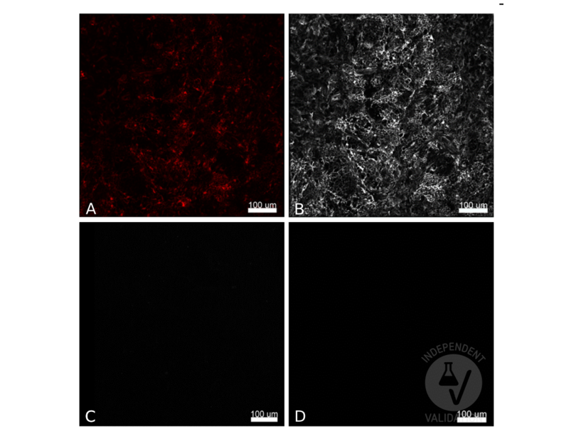
![CX3CL1 positive cells in CX3CL1mcherry murine thymus (A, mCherry) were stained with ABIN1043867 and an AF647 conjugated secondary antibody (B) or with the secondary antibody alone (C). CX3CL1 positive cells in CX3CL1 wt murine thymus were stained with ABIN1043867 and the AF647 conjugated antibody (D). Scale bar 100?m, 20x magnification, 1000 resolution]() CX3CL1 positive cells in CX3CL1mcherry murine thymus (A, mCherry) were stained with ABIN1043867 and an AF647 conjugated secondary antibody (B) or with the secondary antibody alone (C). CX3CL1 positive cells in CX3CL1 wt murine thymus were stained with ABIN1043867 and the AF647 conjugated antibody (D). Scale bar 100?m, 20x magnification, 1000 resolution
Protokoll
CX3CL1 positive cells in CX3CL1mcherry murine thymus (A, mCherry) were stained with ABIN1043867 and an AF647 conjugated secondary antibody (B) or with the secondary antibody alone (C). CX3CL1 positive cells in CX3CL1 wt murine thymus were stained with ABIN1043867 and the AF647 conjugated antibody (D). Scale bar 100?m, 20x magnification, 1000 resolution
Protokoll -
- Format
- Liquid
- Konzentration
- 1.16 mg/mL
- Buffer
- 0.02 M Potassium Phosphate, 0.15 M Sodium Chloride, pH 7.2, 0.01% (w/v) Sodium Azide
- Konservierungsmittel
- Sodium azide
- Vorsichtsmaßnahmen
- This product contains sodium azide: a POISONOUS AND HAZARDOUS SUBSTANCE which should be handled by trained staff only.
- Handhabung
- Avoid cycles of freezing and thawing.
- Lagerung
- 4 °C/-20 °C
- Informationen zur Lagerung
- Store vial at -20° C prior to opening. Aliquot contents and freeze at -20° C or below for extended storage. Centrifuge product if not completely clear after standing at room temperature. This product is stable for several weeks at 4° C as an undiluted liquid. Dilute only prior to immediate use.
- Haltbarkeit
- 12 months
-
-
: "Injury primes mutation-bearing astrocytes for dedifferentiation in later life." in: Current biology : CB, (2023) (PubMed).
: "Diet suppresses glioblastoma initiation in mice by maintaining quiescence of mutation-bearing neural stem cells." in: Developmental cell, (2023) (PubMed).
: "Molecular sensing of mechano- and ligand-dependent adhesion GPCR dissociation." in: Nature, Vol. 615, Issue 7954, pp. 945-953, (2023) (PubMed).
: "Overexpression of Lin28A in neural progenitor cells in vivo does not lead to brain tumor formation but results in reduced spine density." in: Acta neuropathologica communications, Vol. 9, Issue 1, pp. 185, (2022) (PubMed).
: "A versatile viral toolkit for functional discovery in the nervous system. ..." in: Cell reports methods, Vol. 2, Issue 6, pp. 100225, (2022) (PubMed).
: "Axon guidance at the spinal cord midline-A live imaging perspective." in: The Journal of comparative neurology, (2021) (PubMed).
: "Extrinsic Regulators of mRNA Translation in Developing Brain: Story of WNTs." in: Cells, Vol. 10, Issue 2, (2021) (PubMed).
: "Co-activation of Sonic hedgehog and Wnt signaling in murine retinal precursor cells drives ocular lesions with features of intraocular medulloepithelioma." in: Oncogenesis, Vol. 10, Issue 11, pp. 78, (2021) (PubMed).
: "The white matter is a pro-differentiative niche for glioblastoma." in: Nature communications, Vol. 12, Issue 1, pp. 2184, (2021) (PubMed).
: "Antinociceptive modulation by the adhesion GPCR CIRL promotes mechanosensory signal discrimination." in: eLife, Vol. 9, (2021) (PubMed).
: "Reticular Fibroblasts Expressing the Transcription Factor WT1 Define a Stromal Niche that Maintains and Replenishes Splenic Red Pulp Macrophages." in: Immunity, Vol. 53, Issue 1, pp. 127-142.e7, (2020) (PubMed).
: "Transcriptional activities of human elongation factor-1α and cytomegalovirus promoter in transgenic dogs generated by somatic cell nuclear transfer." in: PLoS ONE, Vol. 15, Issue 6, pp. e0233784, (2020) (PubMed).
: "Gastric squamous-columnar junction contains a large pool of cancer-prone immature osteopontin responsive Lgr5-CD44+ cells." in: Nature communications, Vol. 11, Issue 1, pp. 84, (2020) (PubMed).
: "Yorkie and JNK revert syncytial muscles into myoblasts during Org-1-dependent lineage reprogramming." in: The Journal of cell biology, Vol. 218, Issue 11, pp. 3572-3582, (2020) (PubMed).
: "Evolution of Ovipositor Length in Drosophila suzukii Is Driven by Enhanced Cell Size Expansion and Anisotropic Tissue Reorganization." in: Current biology : CB, Vol. 29, Issue 12, pp. 2075-2082.e6, (2020) (PubMed).
: "Disynaptic cerebrocerebellar pathways originating from multiple functionally distinct cortical areas." in: eLife, Vol. 9, (2020) (PubMed).
: "Cellular and molecular properties of neural progenitors in the developing mammalian hypothalamus." in: Nature communications, Vol. 11, Issue 1, pp. 4063, (2020) (PubMed).
: "From 2D to 3D: Promising Advances in Imaging Lung Structure." in: Frontiers in medicine, Vol. 7, pp. 343, (2020) (PubMed).
: "Osterix-Cre marks distinct subsets of CD45- and CD45+ stromal populations in extra-skeletal tumors with pro-tumorigenic characteristics." in: eLife, Vol. 9, (2020) (PubMed).
: "Genetic background mutations drive neural circuit hyperconnectivity in a fragile X syndrome model." in: BMC biology, Vol. 18, Issue 1, pp. 94, (2020) (PubMed).
-
: "Injury primes mutation-bearing astrocytes for dedifferentiation in later life." in: Current biology : CB, (2023) (PubMed).
-
- Target
- RFP (Red Fluorescent Protein (RFP))
- Andere Bezeichnung
- RFP (RFP Produkte)
- Hintergrund
-
Synonyms: DsRed, rDsRed, Discosoma sp. Red Fluorescent Protein, Red fluorescent protein drFP583
Fluorescent proteins such as Discosoma Red Fluorescent Protein (DsRed) from sea anemone Discosoma sp. mushroom or green fluorescent protein (GFP) from Aequorea victoria jellyfish are widely used in research practice. Fusion RFP and GFP commonly serve as marker for gene expression and protein localization. As DsRed and GFP share only 19% identity, therefore, in general, anti-GFP antibodies do not recognize DsRed protein and vice versa. Structurally, Discosoma red fluorescent protein is similar to Aequorea green fluorescent protein in terms of its overall fold (a β-can) and chromophore-formation chemistry. However, Discosoma red fluorescent protein undergoes an additional step in the chromophore maturation and obligates tetrameric structure.
-


