MYCN Antikörper (AA 401-464)
 (1 reference)
 (1 Validierung)
Kurzübersicht für MYCN Antikörper (AA 401-464) (ABIN760676)
Target
Alle MYCN Antikörper anzeigenReaktivität
Wirt
Klonalität
Konjugat
Applikation
-
-
Bindungsspezifität
- AA 401-464
-
Verwendungszweck
- n-Myc Polyclonal Antibody
-
Kreuzreaktivität
- Human, Maus
-
Homologie
- Rat,Cow,Pig
-
Aufreinigung
- Purified by Protein A.
-
Immunogen
- KLH conjugated synthetic peptide derived from human n-Myc
-
Isotyp
- IgG
-
-
anti-N-Myc Proto-Oncogene Protein (MYCN) (AA 168-267) antibody
MYCN Reaktivität: Human WB, IHC, IF Wirt: Kaninchen Polyclonal unconjugated
anti-N-Myc Proto-Oncogene Protein (MYCN) (AA 337-464) antibodyMYCN Reaktivität: Human ELISA, IHC, FACS Wirt: Maus Monoclonal 2A4E9 unconjugated
anti-N-Myc Proto-Oncogene Protein (MYCN) (AA 1-100) antibodyMYCN Reaktivität: Human WB, ELISA, RNAi Wirt: Maus Monoclonal 3H4 unconjugated
anti-N-Myc Proto-Oncogene Protein (MYCN) (AA 100-150) antibodyMYCN Reaktivität: Human, Maus WB, ICC, IP Wirt: Kaninchen Monoclonal BLR353N unconjugated Recombinant Antibody
anti-N-Myc Proto-Oncogene Protein (MYCN) (AA 322-351), (C-Term) antibodyMYCN Reaktivität: Human WB, IHC (p) Wirt: Kaninchen Polyclonal RB18771 unconjugated
anti-N-Myc Proto-Oncogene Protein (MYCN) (Internal Region) antibodyMYCN Reaktivität: Human, Maus, Ratte WB, IHC, IF, ICC Wirt: Kaninchen Polyclonal unconjugated
anti-N-Myc Proto-Oncogene Protein (MYCN) (C-Term) antibodyMYCN Reaktivität: Human WB, ChIP-seq Wirt: Kaninchen Polyclonal unconjugated
anti-N-Myc Proto-Oncogene Protein (MYCN) antibodyMYCN Reaktivität: Human, Maus, Ratte IHC Wirt: Kaninchen Polyclonal unconjugated
anti-N-Myc Proto-Oncogene Protein (MYCN) antibodyMYCN Reaktivität: Human, Maus, Ratte ELISA, IHC Wirt: Kaninchen Polyclonal unconjugated
anti-N-Myc Proto-Oncogene Protein (MYCN) (AA 1-464) antibodyMYCN Reaktivität: Human WB Wirt: Kaninchen Polyclonal unconjugated
-
-
Applikationshinweise
- WB(), IHC-P(1:200-400)
-
Beschränkungen
- Nur für Forschungszwecke einsetzbar
-
-
- by
- Alamo Laboratories Inc
- No.
- #029800
- Datum
- 21.08.2014
- Antigen
- Chargennummer
- 120917
- Validierte Anwendung
- Western Blotting
- Positivkontrolle
- MCF7 cells (variant MCF7/VP)00284-0/pdf)
- Negativkontrolle
- SK-N-SH cells - RNA
- Bewertung
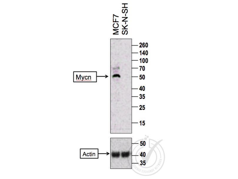
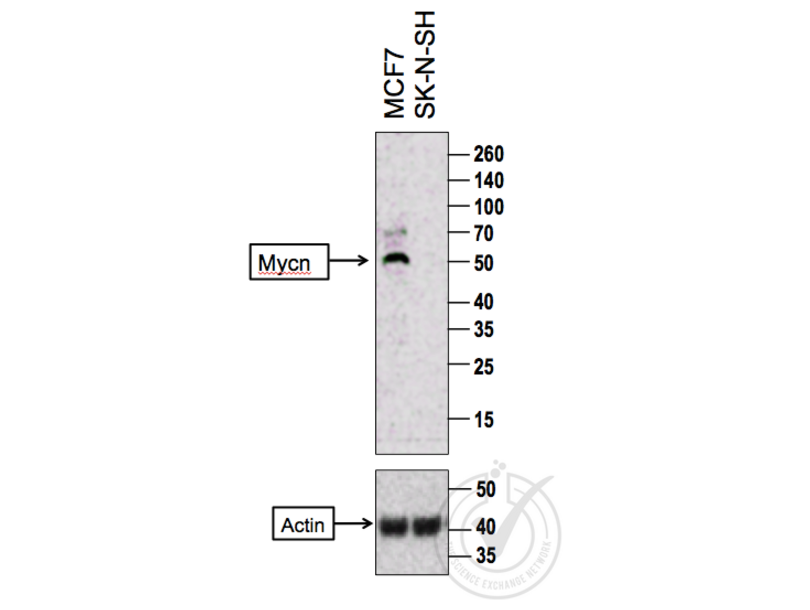
- A strong band was observed in the positive control sample at the correct molecular weight. An additional band was also observed in the positive sample at a higher molecular weight which may represent non-specific binding. No bands were observed in the negative control sample.
- Primärantikörper
- Antigen: Mycn (MYCN)
- Catalog number: ABIN760676
- Lot number: 120917
- Antibody Dilution: 1:200
- Sekundärantikörper
- Antigen: Goat Anti-Rabbit IgG (H + L)-HRP Conjugate
- Lot number: L170-6515
- Antibody Dilution: 1:10,000
- Full Protocol
- 1. The cell extracts were heated at 95°C for 5 minutes in 1X SDS Sample Buffer containing 1% SDS and 1.25% β-mercaptoethanol.
- 2. 15 μl of heated culture-media were loaded and resolved on 8-16% SDS-polyacrylamide gel.
- 3. The Thermo Scientific - Spectra Multicolor Broad Range (Cat # 26634) were used as molecular mass markers.
- 4. Proteins were then transferred onto PVDF membrane by wet transfer and protein transfer was confirmed with Ponceau-S staining.
- 5. The PVDF membrane was incubated with 25 ml of blocking buffer [Tris Buffered Saline, pH 7.4 plus 0.1% TW20 (TBST)] containing 5% (W/V) BSA at room temperature for 1 hour.
- 6. The membrane was rinsed with TBST once.
- 7. The membrane was immersed with the protein side up in the primary antibody solution in TBST containing 5% (W/V) BSA and incubated for 13 hours at 4°C.
- 8. The membrane was rinsed in TBST thrice for 5 minutes each.
- 9. The membrane was incubated in the HRP-conjugated secondary antibody solution in TBST containing 5% (W/V) BSA and incubated for 1 hour at room temperature (~26°C) with gentle agitation.
- 10. The membrane was rinsed thrice TBST thrice for 5 minutes each.
- 11. The membrane was rinsed in TBS twice for 30 seconds each.
- 12. Signals were detected with ECL-2 Substrate. The blot was scanned for 45 minutes.
- 13. The membrane was rinsed three times TBST.
- 14. Incubated in Acidic Glycine Stripping Buffer at room temperature with gentle agitation for 3 times, 10 minutes each.
- 15. The membrane was washed in TBST 2 times for 10 minutes each.
- 16. Repeated Steps 5-12 with the loading control antibody (for Anti-actin) and its matching secondary antibody.
- Anmerkungen
- - No experimental challenges noted.
Validierung #029800 (Western Blotting)
 Validierungsbilder
![Figure 1: Western Blot for MYCN. Arrowhead indicates the expected molecular weight of ~49 kDa.]() Figure 1: Western Blot for MYCN. Arrowhead indicates the expected molecular weight of ~49 kDa.
Protokoll
Figure 1: Western Blot for MYCN. Arrowhead indicates the expected molecular weight of ~49 kDa.
Protokoll -
-
Format
- Liquid
-
Konzentration
- 1 μg/μL
-
Buffer
- 0.01M TBS( pH 7.4) with 1 % BSA, 0.02 % Proclin300 and 50 % Glycerol.
-
Konservierungsmittel
- ProClin
-
Vorsichtsmaßnahmen
- This product contains ProClin: a POISONOUS AND HAZARDOUS SUBSTANCE which should be handled by trained staff only.
-
Lagerung
- 4 °C,-20 °C
-
Informationen zur Lagerung
- Shipped at 4°C. Store at -20°C for one year. Avoid repeated freeze/thaw cycles.
-
Haltbarkeit
- 12 months
-
-
-
: "Serum-circulating miRNAs predict neuroblastoma progression in mouse model of high-risk metastatic disease." in: Oncotarget, Vol. 7, Issue 14, pp. 18605-19, (2016) (PubMed).
-
-
- MYCN (N-Myc Proto-Oncogene Protein (MYCN))
-
Andere Bezeichnung
- N-Myc
-
Hintergrund
-
Synonyms: NMYC, ODED, MODED, N-myc, bHLHe37, N-myc proto-oncogene protein, Class E basic helix-loop-helix protein 37, MYCN
Background: This gene is a member of the MYC family and encodes a protein with a basic helix-loop-helix (bHLH) domain. This protein is located in the nucleus and must dimerize with another bHLH protein in order to bind DNA. Amplification of this gene is associated with a variety of tumors, most notably neuroblastomas. [provided by RefSeq, Jul 2008].
-
Gen-ID
- 4613
-
UniProt
- P04198
Target
-