Histone 3 Antikörper (H3K27ac)
 (1 Validierung)
Kurzübersicht für Histone 3 Antikörper (H3K27ac) (ABIN2667903)
Target
Alle Histone 3 (H3) Antikörper anzeigenReaktivität
Wirt
Klonalität
Konjugat
Applikation
-
-
Bindungsspezifität
- H3K27ac
-
Verwendungszweck
- Histone H3K27ac antibody (pAb), sample
-
Aufreinigung
- Protein A Chromatography
-
Immunogen
- This Histone H3 acetyl Lys27 antibody was raised against a peptide including acetyl-lysine 27 of histone H3.
-
Isotyp
- IgG
-
-
anti-Histone H3 (H3) (3meLys4) antibody
H3 Reaktivität: Human, Saccharomyces cerevisiae WB, IF, ChIP, ICC, DB, ChIP-seq, CUT&Tag, CUT&RUN Wirt: Kaninchen Polyclonal unconjugated
anti-Histone H3 (H3) (H3K4me) antibodyH3 Reaktivität: Human WB, IF, ChIP, IP, ChIP-seq, CUT&RUN Wirt: Kaninchen Polyclonal unconjugated
anti-Histone H3 (H3) (H3K27ac) antibodyH3 Reaktivität: Human, Saccharomyces cerevisiae WB, IF, IHC, ChIP, ICC, DB, ChIP-seq, CUT&Tag Wirt: Kaninchen Polyclonal unconjugated
anti-Histone H3 (H3) (H3K9ac) antibodyH3 Reaktivität: Human, Maus WB, IF, ChIP, ICC, DB, ChIP-seq, CUT&Tag Wirt: Kaninchen Polyclonal unconjugated
anti-Histone H3 (H3) (H3K36me3) antibodyH3 Reaktivität: Human, Maus WB, IF, ChIP, DB, ChIP-seq Wirt: Maus Monoclonal MABI 0333 unconjugated
anti-Histone H3 (H3) (H3K27me) antibodyH3 Reaktivität: Human WB, IF, IHC, ChIP, IP, ChIP-seq Wirt: Kaninchen Polyclonal unconjugated
anti-Histone H3 (H3) (AA 71-136) antibodyH3 Reaktivität: Human WB, FACS, IHC (p), IF (cc), IF (p), IHC (fro) Wirt: Kaninchen Polyclonal unconjugated
anti-Histone H3 (H3) (H3R17me) antibodyH3 Reaktivität: Human WB, IF, IP Wirt: Kaninchen Polyclonal unconjugated
anti-Histone H3 (H3) (H3K36me3) antibodyH3 Reaktivität: Human WB, IF, IHC, ChIP, IP, ChIP-seq Wirt: Kaninchen Polyclonal unconjugated
anti-Histone H3 (H3) (H3K36me2) antibodyH3 Reaktivität: Human WB, IF, IHC, ChIP, IP, ChIP-seq Wirt: Kaninchen Polyclonal unconjugated
-
-
Applikationshinweise
- ChIP: 10 μg per ChIP ChIP-Seq: 5 μg each ICC/IF: 1 - 5 μg/mL dilution WB*: 0.1 - 1 μg/mL dilution CUT&Tag: 1 μg per 50 µL reaction* *This antibody has been validated for CUT&Tag using Active Motif's CUT&Tag-IT Assay Kit, Catalog No. 53160. *Note: many chromatin-bound proteins are not soluble in a low salt nuclear extract and fractionate to the pellet. Therefore, we recommend a High Salt / Sonication Protocol when preparing nuclear extracts for Western blot. modENCODE validation: this antibody was validated for ChIP-Seq in this study (see references). NGS-QC certification: this antibody has been processed by the NGS-QC generator.
-
Beschränkungen
- Nur für Forschungszwecke einsetzbar
-
-
- by
- Gianluca Zambanini, Anna Nordin and Claudio Cantù; Cantù Lab, Gene Regulation during Development and Disease, Linköping University
- No.
- #104485
- Datum
- 09.06.2022
- Antigen
- H3K27ac
- Chargennummer
- 6921014
- Validierte Anwendung
- Cleavage Under Targets and Release Using Nuclease
- Positivkontrolle
Polyclonal rabbit anti-H3K4me (antibodies-online, ABIN3023251)
- Negativkontrolle
Polyclonal guinea pig anti-rabbit IgG (antibodies-online, ABIN101961)
- Bewertung
Passed. ABIN2667903 allows for H3K27Ac targeted digestion using CUT&RUN in mouse fore limb (11.5) cells.
- Primärantikörper
- ABIN2667903
- Sekundärantikörper
- Full Protocol
- Cell harvest and nuclear extraction
- Dissect 3 Fore limbs (11.5 DAC) from mouse strain RjOrl:SWISS for each sample.
- Dissociate the tissue into single cells in TrypLE for 15 min at 37 °C.
- Centrifuge cell solution 5 min at 800 x g at RT.
- Remove the liquid carefully.
- Gently resuspend cells in 1 mL of Nuclear Extraction Buffer (20 mM HEPES-KOH pH 8.2, 20% Glycerol, 0.05% IGEPAL, 0.5 mM Spermidine, 10 mM KCl, Roche Complete Protease Inhibitor EDTA-free).
- Move the solution to a 2 mL centrifuge tube.
- Pellet the nuclei 800 x g for 5 min.
- Repeat the NE wash twice for a total of three washes.
- Resuspend the nuclei in 20 µL NE Buffer per sample.
- Concanavalin A beads preparation
- Prepare one 2 mL microcentrifuge tube.
- Gently resuspend the magnetic Concanavalin A Beads (antibodies-online, ABIN6952467).
- Pipette 20 µL Con A Beads slurry for each sample into the 2 mL microcentrifuge tube.
- Place the tube on a magnet stand until the fluid is clear. Remove the liquid carefully.
- Remove the microcentrifuge tube from the magnetic stand.
- Pipette 1 mL Binding Buffer (20 mM HEPES pH 7.5, 10 mM KCl, 1 mM CaCl2, 1 mM MnCl2) into the tube and resuspend ConA beads by gentle pipetting.
- Spin down the liquid from the lid with a quick pulse in a table-top centrifuge.
- Place the tubes on a magnet stand until the fluid is clear. Remove the liquid carefully.
- Remove the microcentrifuge tube from the magnetic stand.
- Repeat the wash twice for a total of three washes.
- Gently resuspend the ConA Beads in a volume of Binding Buffer corresponding to the original volume of bead slurry, i.e. 20 µL per sample.
- Nuclei immobilization – binding to Concanavalin A beads
- Carefully vortex the nuclei suspension and add 20 µL of the Con A beads in Binding Buffer to the cell suspension for each sample.
- Close tube tightly incubates 10 min at 4 °C.
- Put the 2 mL tube on the magnet stand and when the liquid is clear remove the supernatant.
- Resuspend the beads in 1 mL of EDTA Wash buffer (20 mM HEPES pH 7.5, 150 mM NaCl, 0.5 mM Spermidine, Roche Complete Protease Inhibitor EDTA-free, 2mM EDTA).
- Incubate 5 min at RT.
- Place the tube on the magnet stand and when the liquid is clear remove the supernatant.
- Resuspend the beads in 200 µl of Wash Buffer (20 mM HEPES pH 7.5, 150 mM NaCl, 0.5 mM Spermidine, Roche Complete Protease Inhibitor EDTA-free) per sample.
- Primary antibody binding
- Divide nuclei suspension into separate 200 µL PCR tubes, one for each antibody (150,000 cells per sample).
- Add 2 µL antibody (anti-H3K27ac antibody ABIN2667903, anti-H3K4me positive control antibody ABIN3023251, and guinea pig anti-rabbit IgG negative control antibody ABIN101961) to the respective tube, corresponding to a 1:100 dilution.
- Incubate at 4 °C ON.
- Place the tubes on a magnet stand until the fluid is clear. Remove the liquid carefully.
- Remove the microcentrifuge tubes from the magnetic stand.
- Wash with 200 µL of Wash Buffer using a multichannel pipette to accelerate the process.
- Repeat the wash five times for a total of six washes.
- pAG-MNase Binding
- Prepare a 1.5 mL microcentrifuge tube containing 100 µL of pAG mix per sample (100 µL of wash buffer + 58.5 µg pAG-MNase per sample).
- Place the PCR tubes with the sample on a magnet stand until the fluid is clear. Remove the liquid carefully.
- Remove tubes from the magnetic stand.
- Resuspend the beads in 100 µL of pAG-MNase premix.
- Incubate 30 min at 4 °C.
- Place the tubes on a magnet stand until the fluid is clear. Remove the liquid carefully.
- Remove the microcentrifuge tubes from the magnetic stand.
- Wash with 200 µL of Wash Buffer using a multichannel pipette to accelerate the process.
- Repeat the wash five times for a total of six washes.
- Resuspend in 100 µL of Wash Buffer.
- MNase digestion and release of pAG-MNase-antibody-chromatin complexes
- Place PCR tubes on ice and allow to chill.
- Prepare a 1.5 mL microcentrifuge tube with 102 µl of 2 mM CaCl2 mix per sample (100 µl Wash Buffer + 2 µL 100 mM CaCl2) and let it chill on ice.
- Always in ice, place the samples on the magnetic rack and when the liquid is clear remove the supernatant.
- Resuspend the samples in 100 µl of the 2 mM CaCl2 mix and incubate in ice for exactly 30 min.
- Place the sample on the magnet stand and when the liquid is clear remove the supernatant.
- Resuspend the sample in 50 µl of 1x Urea STOP Buffer (8.5 M Urea, 100 mM NaCl, 2 mM EGTA, 2 mM EDTA, 0.5% IGEPAL).
- Incubate the samples 1h at 4°C.
- Transfer the supernatant containing the pAG-MNase-bound digested chromatin fragments to fresh 200 µl PCR tubes.
- DNA Clean up
- Take the Mag-Bind® TotalPure NGS beads (Omega Bio-Tek, M1378-01) from the storage and wait until they are at RT.
- Add 2x volume of beads to each sample (e.g. 100 µL of beads for 50 µL of sample).
- Incubate the beads and the sample for 15 min at RT.
- During incubation prepare fresh EtOH 80%.
- Place the PCR tubes on a magnet stand and when the liquid is clear remove the supernatant.
- Add 200 µl of fresh 80% EtOH to the sample without disturbing the beads (Important!!! Do NOT resuspend the beads or remove the tubes from the magnet stand or the sample will be lost).
- Incubate 30 sec at RT.
- Remove the EtOH from the sample.
- Repeat the wash with 80% EtOH.
- Resuspend the beads in 25 µL of 10 mM Tris-HCl pH 8.2.
- Incubate the sample for 2 min at RT.
- Repeat the 2x beads clean up as described before (this time with 50 µL of beads for each sample).
- Resuspend the beads + DNA in 20 µL of 10 mM Tris-HCl pH 8.2.
- Library preparation and sequencing
- Prepare Libraries using KAPA HyperPrep Kit using KAPA Dual-Indexed adapters according to protocol.
- Sequence samples on an Illumina NextSeq 500 sequencer, using a NextSeq 500/550 High Output Kit v2.5 (75 Cycles), 36 bp PE.
- Peak calling
- Trim reads using using bbTools bbduk (BBMap - Bushnell B. - sourceforge.net/projects/bbmap/) to remove adapters, artifacts and repeat sequences.
- Map aligned reads to the hg38 human genome using bowtie with options -m 1 -v 0 -I 0 -X 500.
- Use SAMtools to convert SAM files to BAM files and remove duplicates.
- Use BEDtools genomecov to produce Bedgraph files.
- Call peaks using SEACR with a 0.001 threshold and the option norm stringent.
- Anmerkungen
The protocol is published in PMID 36355069
.
Validierung #104485 (Cleavage Under Targets and Release Using Nuclease)
 Validierungsbilder
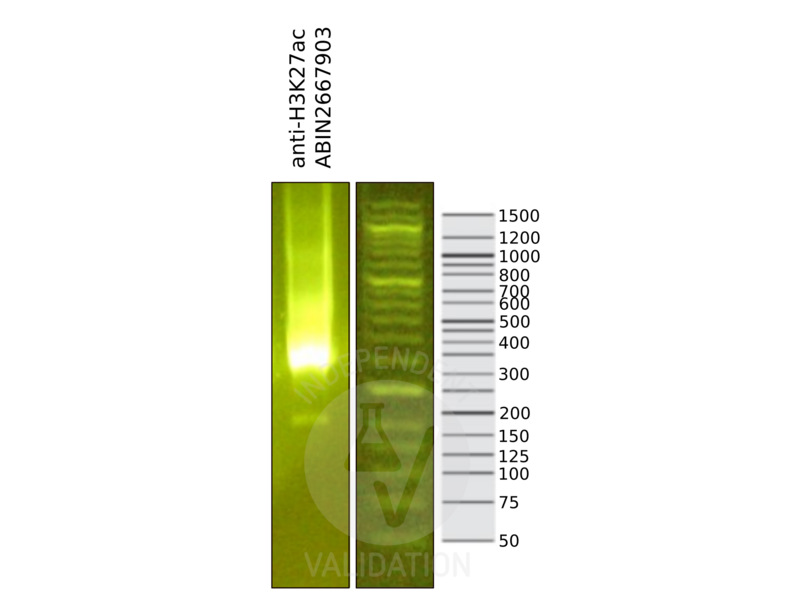
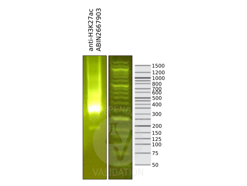
![Library profiles comparing fragment size distributions on an E-Gel EX 2% agarose gel (Thermo Fisher). Fragments obtained from CUT&RUN using anti-H3K27ac antibody ABIN2667903 after library preparation, compared to the E-Gel Sizing DNA Ladder (Thermo Fisher).]() Library profiles comparing fragment size distributions on an E-Gel EX 2% agarose gel (Thermo Fisher). Fragments obtained from CUT&RUN using anti-H3K27ac antibody ABIN2667903 after library preparation, compared to the E-Gel Sizing DNA Ladder (Thermo Fisher).
Library profiles comparing fragment size distributions on an E-Gel EX 2% agarose gel (Thermo Fisher). Fragments obtained from CUT&RUN using anti-H3K27ac antibody ABIN2667903 after library preparation, compared to the E-Gel Sizing DNA Ladder (Thermo Fisher).
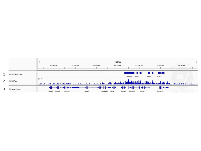
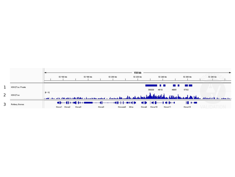
![1. Peaks called by SEACR from CUT&RUN data using anti- H3K27ac antibody ABIN2667903. 2. Alignment tracks from CUT&RUN targeting H3K27ac in mouse fore limb (11.5) cells using anti-H3K27ac antibody ABIN2667903 showing the Hoxa locus 3. RefSeq Genes.]() 1. Peaks called by SEACR from CUT&RUN data using anti- H3K27ac antibody ABIN2667903. 2. Alignment tracks from CUT&RUN targeting H3K27ac in mouse fore limb (11.5) cells using anti-H3K27ac antibody ABIN2667903 showing the Hoxa locus 3. RefSeq Genes.
Protokoll
1. Peaks called by SEACR from CUT&RUN data using anti- H3K27ac antibody ABIN2667903. 2. Alignment tracks from CUT&RUN targeting H3K27ac in mouse fore limb (11.5) cells using anti-H3K27ac antibody ABIN2667903 showing the Hoxa locus 3. RefSeq Genes.
Protokoll -
-
Konzentration
- 1 μg/μL
-
Buffer
- Purified IgG in PBS with 30 % glycerol and 0.035 % sodium azide.
-
Konservierungsmittel
- Sodium azide
-
Vorsichtsmaßnahmen
- This product contains Sodium azide: a POISONOUS AND HAZARDOUS SUBSTANCE which should be handled by trained staff only.
-
Handhabung
- Avoid repeated freeze/thaw cycles by aliquoting items into single-use fractions,Keep all reagents on ice when not in storage
-
Lagerung
- -20 °C
-
Informationen zur Lagerung
- Some products may be shipped at room temperature. This will not affect their stability or performance. Avoid repeated freeze/thaw cycles by aliquoting items into single-use fractions for storage at -20°C for up to 2 years. Keep all reagents on ice when not in storage.
-
Haltbarkeit
- 24 months
-
-
- Histone 3 (H3) (Histone H3 (H3))
-
Andere Bezeichnung
- Histone H3
-
Hintergrund
- Histone H3 is one of the core components of the nucleosome. The nucleosome is the smallest subunit of chromatin and consists of 147 base pairs of DNA wrapped around an octamer of core histone proteins (two each of Histone H2A, Histone H2B, Histone H3 and Histone H4). Histone H1 is a linker histone, present at the interface between the nucleosome core and DNA entry/exit points, it is responsible for establishing higher-order chromatin structure. Chromatin is subject to a variety of chemical modifications, including post-translational modifications of the histone proteins and the methylation of cytosine residues in the DNA. Reported histone modifications include acetylation, methylation, phosphorylation, ubiquitylation, glycosylation, ADP-ribosylation, carbonylation and SUMOylation, they play a major role in regulating gene expression. Lysine N-e-acetylation is a dynamic, reversible and tightly regulated protein and histone modification that plays a major role in chromatin remodeling and in the regulation of gene expression in various cellular functions. Acetylation of histone H3 occurs at several different lysine positions in the histone tail, and is performed by Histone Acetyltransferases (HATs) such as CBP/p300. Acetylation of histone H3 at Lys27 is associated with transcriptional activation. Histone H3K27 can also be mono-, di- or trimethylated by different histone methyltransferases, such as EZH2 or NSD3. While histone methylation can be associated with transcriptional activation or repression, methylation of Lysine 27 of histone H3 is mainly associated with transcriptional repression.
-
Molekulargewicht
- 17 kDa
-
NCBI Accession
- NP_003522
Target
-