Rhinovirus Antikörper
 (1 Validierung)
Kurzübersicht für Rhinovirus Antikörper (ABIN5688681)
Target
Reaktivität
Wirt
Klonalität
Applikation
Klon
-
-
Spezifität
- Specific for the VP3 of numerous Rhino types. Does not cross react with Enteroviruses, Influenza A, Influenza B, RSV, Adenovirus, Para 1, Para 2, Para 3, Chlamydia pneumoniae, Rhinovirus pneumoniae & Hepatitis A virus.
-
Aufreinigung
- Rhinovirus Antibody is purified from ascites fluid or culture medium by protein A chromoatography or sequential differential precipitations.
-
Isotyp
- IgG1
-
-
-
Applikationshinweise
- Rhinovirus Antibody [4966] can be used at 1:10-1:50 for Western blotting, Immunohistochemistry, and Immunoflorescence. For ELISA use at 1:20-1:200.
-
Beschränkungen
- Nur für Forschungszwecke einsetzbar
-
-
- by
- Virology Research Lab, Department of Pathology and Laboratory Medicine, Faculty of Medicine, University of British Columbia
- No.
- #103804
- Datum
- 14.06.2019
- Antigen
- Rhinovirus
- Chargennummer
- 22523-1902
- Validierte Anwendung
- Immunohistochemistry
- Positivkontrolle
Human rhinovirus 14 infected tisssue (2moi/cell)
- Negativkontrolle
Uninfected tissue
no primary antibody controls
- Bewertung
The rhinovirus antibody ABIN5688681 successfully labels the targeted antigen in a 3D respiratory tissue model in IHC.
- Primärantikörper
- ABIN5688681
- Sekundärantikörper
- Goat Anti-Mouse IgG H&L AF488 antibody (Abcam, ab150117)
- Full Protocol
- Grow EpiAirway 3D tissue model (MatTek) in Normal Human Bronchial Epithelial Cells - Growth Medium MatTek, AIR-100-ASY-AFAB) at 37°C and 5% CO2.
- Infect tissue model with HRV-14 (ATCC, VR284) at 2MOI/cell).
- Incubate for 72h at 34°C and 5% CO2.
- Fix tissue in 4% buffered formalin for 45 min at 23°C.
- Rinse sections 2x for 2min with PBS.
- Permeabilized tissues with 0.5% saponin for 15min,rinsed 2X with PBS.
- Block tissue in PBS containing 5% BSA (R&D Systems, DY995) for 1h at 4°C.
- Blot excess serum from sections.
- Incubate tissue with primary mouse anti-rhinovirus antibody (antibody-online, ABIN5688681, lot 22523-1902)diluted 1:50 in PBS containing 1% BSA and 0.2% tween 20 ON at 4°C. Includeno primaryantibody negative controls.
- Rinse tissue 3x with PBS. Keep negative controls in a separate container.
- Incubate sections with secondary Goat Anti-Mouse IgG H&L AF488 antibody (Abcam, ab150117)diluted 1:250x in PBS containing 1% BSA for 1h at 23°C.
- Rinsed sections 3x with PBS.
- Mount sections in fluoroshield mounting medium with DAPI (Abcam, ab104139).
- Acquire images with Zeiss Axioplan 2 Imaging Microscope 20X objective, and analyze usingAxio Vision software.
- Anmerkungen
ABIN5688681 showed some signals only in an HRV-14 infected human 3D tissue model that had not undergone antigen retrieval. In HRV-14 samples that had been incubated for 15min in citrate buffer pH5.0 at sub-boiling temperatures no rhinovirus was detected. In order to block unspecific binding of the antibody these samples were incubated ON at 4°C in blocking buffer. It is possible that the blocking step was too long, thus preventing the antibody from binding to the antigen.
Omitting the antigen retrieval step and reducing the blocking time resulted in a specific signal in the rhinovirus infected samples. However, unspecific background signal was also visible in the uninfected negative controls. It may be possible to reduce the background signal by increasing the incubation during the blocking step.
Validierung #103804 (Immunohistochemistry)
 Validierungsbilder
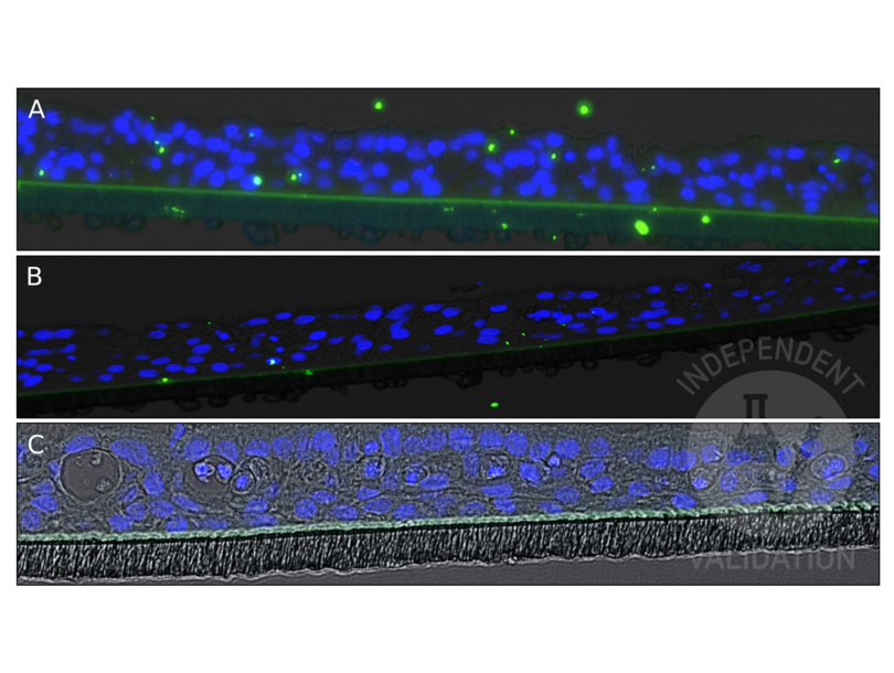
![Staining of rhinovirus in a human 3D respiratory tissue model infected with HRV-14 using ABIN5688681 and an AF488-conjugated secondary antibody (A). In comparison, the same tissue model without HRV-14 infection (B) or stained using the secondary antibody only (C). Stained rhinovirus appears as a green signal, DAPI counterstain in blue.]() Staining of rhinovirus in a human 3D respiratory tissue model infected with HRV-14 using ABIN5688681 and an AF488-conjugated secondary antibody (A). In comparison, the same tissue model without HRV-14 infection (B) or stained using the secondary antibody only (C). Stained rhinovirus appears as a green signal, DAPI counterstain in blue.
Protokoll
Staining of rhinovirus in a human 3D respiratory tissue model infected with HRV-14 using ABIN5688681 and an AF488-conjugated secondary antibody (A). In comparison, the same tissue model without HRV-14 infection (B) or stained using the secondary antibody only (C). Stained rhinovirus appears as a green signal, DAPI counterstain in blue.
Protokoll -
-
Format
- Liquid
-
Buffer
- Rhinovirus antibody is in a phosphate saline buffer (0.01M, pH 7.2) containing 0.1 % sodium azide preservative. No stabilizing proteins have been added.
-
Konservierungsmittel
- Sodium azide
-
Vorsichtsmaßnahmen
- This product contains Sodium azide: a POISONOUS AND HAZARDOUS SUBSTANCE which should be handled by trained staff only.
-
Lagerung
- -20 °C
-
Informationen zur Lagerung
- Rhinovirus antibody can be stored at -20°C, stable for one year.
-
-
- Rhinovirus
-
Substanzklasse
- Virus
Target
-


