COL1A2/COL1A1 Antikörper
 (62 Referenzen)
 (2 validations)
-
- Highlights
-
- High quality Primärantikörper for the detection of .
- Reliable product with high standard validation data
- Target
- COL1A2/COL1A1
- Reaktivität
- Human, Rind (Kuh), Maus, Schwein, Ratte
-
Wirt
- Kaninchen
-
Klonalität
- Polyklonal
-
Konjugat
- Unkonjugiert
- Applikation
- ELISA, Western Blotting (WB), Dot Blot (DB), FLISA, Flow Cytometry (FACS), Fluorescence Microscopy (FM), Immunohistochemistry (IHC), Immunoprecipitation (IP)
- Hersteller Produkt- Nr.
- 600-401-103-0.1
- Hersteller
- Rockland
- Verwendungszweck
- Collagen Type I Antibody
- Kreuzreaktivität (Details)
- Some class-specific anti-collagens may be specific for three-dimensional epitopes which may result in diminished reactivity with denatured collagen or formalin-fixed, paraffin embedded tissues.
- Produktmerkmale
- Synonyms: rabbit anti-collagen type I antibody, Collagen Of Skin Tendon And Bone, Collagen Type 1 antibody, Collagen type I alpha 1 antibody, Collagen alpha-1 (I) chain, Alpha-1 type I collagen, type 1 procollagen alpha 1
- Aufreinigung
- COLLAGEN I Antibody has been prepared by immunoaffinity chromatography using immobilized antigens.
- Sterilität
- Sterile filtered
- Immunogen
-
Immunogen: Collagen Type I from human and bovine placenta
Immunogen Type: Native Protein
- Isotyp
- IgG
- Produktspezifische Information
-
- What can the COL1A2/COL1A1 Antibody ABIN5596819 be used for?
- This polyclonal COL1A2/COL1A1 Antibody detects COL1A2/COL1A1. The COL1A2/COL1A1 Antibody has been validated for various applications and can be used for the detection of COL1A2/COL1A1 and derivatives by ELISA, Western Blotting, Dot Blot, FLISA, Flow Cytometry, Fluorescence Microscopy, Immunohistochemistry, Immunoprecipitation.
- What validation data is available for this COL1A2/COL1A1 Antibody?
- The Primärantikörper is referenced in 62 publications and is characterized by a proven, very high reliability. It has currently 41 product images that show its performance in a variety of applications. The product is currently available in 100 μg, 500 μg quantities. COL1A2/COL1A1 Antibody for the detection of COL1A2/COL1A1 and derivatives.
-
anti-COL1A2/COL1A1 (AA 1200-1217) antibody
Reaktivität: Human ELISA, WB Wirt: Kaninchen Polyclonal unconjugated
-
- Applikationshinweise
-
Flow Cytometry Dilution: User Optimized
Immunohistochemistry Dilution: 1:50 - 1:200
Application Note: Anti-Collagen Type I has been tested by Western blot, dot blot, and IHC and is suitable for indirect trapping ELISA for quantitation of antigen in serum using a standard curve, IP, native PAGE, immunofluorescence, and FC for highly sensitive qualitative analysis.
Western Blot Dilution: 1:1,000 - 1:10,000
Immunoprecipitation Dilution: 1:100
FLISA Dilution: 1:100
ELISA Dilution: 1:5,000 - 1:50,000
IF Microscopy Dilution: User Optimized
Other: User Optimized
- Beschränkungen
- Nur für Forschungszwecke einsetzbar
-
- by
- NanoTemper Technologies
- No.
- #103813
- Datum
- 23.07.2019
- Antigen
- COL1
- Chargennummer
- 41897
- Validierte Anwendung
- Unfolding Profile
- Positivkontrolle
- ABIN5596819
- Negativkontrolle
- Bewertung
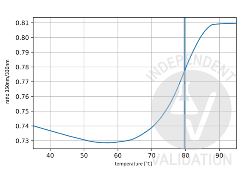
Passed. ABIN5596819 showed Ti at 79.6°C and a clear unfolding profile with one unfolding event. This suggests that the antibody is properly folded and functional.
- Primärantikörper
- Sekundärantikörper
- Full Protocol
- Dilute ABIN5596819 in PBS buffer (Roth, 1058.1, lot 285231988) to get a final volume of 30µl at a concentration of 0.1µM.
- Load sample into Tycho capillary (NanoTemper Technologies, TY-C001).
- Run Tycho measurement.
- Anmerkungen
Tycho is designed to run quick and precise protein quality check experiments. Tycho uses intrinsic protein fluorescence to follow protein unfolding while running a fast thermal ramp, yielding results in 3min. A protein’s unfolding behavior is characterized by various parameters, most notably the inflection temperature (Ti). The Ti can be used to identify properly folded protein, to compare different batches, or to analyze the influence of storage/transport conditions on a protein. An absence of Ti would suggest that the protein is already unfolded and therefore most likely nonfunctional.
Validierung #103813 (Unfolding Profile)
 Validierungsbilder
![Unfolding profile of ABIN5596819. The fluorescence signal is plotted against temperature. The vertical line indicates the Ti at 79.6 °C.]() Unfolding profile of ABIN5596819. The fluorescence signal is plotted against temperature. The vertical line indicates the Ti at 79.6 °C.
Protokoll
Unfolding profile of ABIN5596819. The fluorescence signal is plotted against temperature. The vertical line indicates the Ti at 79.6 °C.
Protokoll -
- by
- MS Validated Antibodies
- No.
- #300048
- Datum
- 07.08.2023
- Antigen
- COL1
- Chargennummer
- 47973
- Validierte Anwendung
- Immunohistochemistry
- Positivkontrolle
Human TMA
Monoclonal rabbit anti-human COL1A2 monoclonal antibody (HL2048)
- Negativkontrolle
- Bewertung
Passed. Staining of collagen type I using ABIN5596819 is consistent with the expected staining pattern.
- Primärantikörper
- ABIN5596819
- Sekundärantikörper
- EnVision Polymer-HRP mouse/rabbit Kit, Dako REAL, K5007
- Full Protocol
- Slide preparation
- Mount 2,5 µm FFPE tissue sections on superfrost slides.
- Deparaffinize tissue sections 3x 5 min in xylene.
- Rehydrate tissue sections in a descending ethanol series for 1 min each 100%, 96%, and 80% ethanol.
- Rinse tissue sections for 5 min in TBST buffer (DAKO, K8000).
- Epitope retrieval
- Autoclave tissue sections for 5 min at 121 °C in 1x Tris-EDTA-citrate buffer pH7.8 (20x Tris-EDTA-citrate buffer stock solution: 5 g Trizma base (Sigma-Aldrich, T1503), 10 g EDTA (Merck, 1.08418), 6.4g tri-sodium citrate (Sigma-Aldrich, C0909), adjust to pH 7.8 using HCL 1 M, ad 1 L with dH2O).
- Rinse tissue sections for 5 min in TBST buffer.
- Peroxidase blocking
- Incubate tissue sections for 10 min in Peroxidase-Blocking Solution (Dako REAL, S2023).
- Rinse tissue sections 2x for 5 min in TBST buffer.
- Antibody incubation
- Dilute primary rabbit anti-collagen type I antibody (antibodies-online, ABIN5596819, lot 47973) diluted 1:15 or 1:50 in antibody diluent (Dako REAL, S2022).
- Cover tissue section with 100-200 µl diluted antibody.
- Incubate tissue sections for 1 h at 37 °C in a moist chamber.
- Rinse tissue sections for 5 min in TBST buffer.
- Apply EnVision Polymer-HRP mouse/rabbit Kit (Dako REAL, K5007) according to manufacturer’s recommendation.
- Rinse tissue sections 2x for 5 min in TBST buffer.
- Staining
- Cover slides for 10 min with DAB-Chromogen (EnVision Polymer-HRP mouse/rabbit Kit, Dako REAL, K5007).
- Wash slides thoroughly with dH2O.
- Counterstain for 15 sec with Hematoxylin (Mayers Hematoxylin: 200ml ddH2O, 0,2g Hematoxylin (Serva, 24420.02), 10 g aluminium potassium sulfate dodecahydrate (Merck, 1.01047), 0,04 g sodium iodate (Merck, 1.06525), 10 g chloral hydrate (Sigma-Aldrich, 15307)).
- Develop for 15 sec in H2O.
- Dehydrate tissue sections in an ascending ethanol series for 1 min each 80%, 96%, 100% ethanol.
- Wash tissue sectiona 3x 5 min in xylene.
- Apply mounting medium and coverslips.
- Image acquisition
- o Acquire images using a Galileo TMAtic (ISENET).
- Anmerkungen
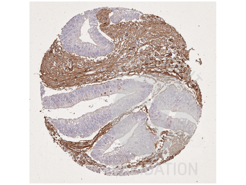
For antibody comparison an antibody test TMA was used that contained 80 normal tissues from 21 different organs and 95 neoplastic tissues from 18 different tumor types. For ABIN5596819 the staining was typically fibrillar. Positive fibrillar structures were often located adjacent to benign and malignant epithelial structures, in smooth muscles or around vessels of all sizes.
The anti-collagen type I antibody ABIN5596819 shows a predominantly fibrillar staining pattern involving stroma components that are often located adjacent to benign and malignant epithelial structures, in smooth muscle or around vessels of all sizes. Collagen I staining is also seen in the stroma of many tumors. The staining of these fibrillar structures by ABIN5596819 is often faint at a dilution of 1:50. Staining of fibrils by ABIN5596819 is stronger at a dilution of 1:15 but in this case a significant background staining occurs in many epithelial tissues. Of note, a membranous staining of intratubular testicular cells was seen by ABIN5596819. Considering the expected staining pattern of an anti-collagen type I antibody, this may represent a cross-reactivity of ABIN5596819.
According to the rather ubiquitous nature of collagen I expression, orthogonal validation is not optimally suited for the validation of collagen I antibodies. In agreement with RNA screening studies, including the Human Protein Atlas (HPA) RNA-seq tissue dataset, the FANTOM5 project, and the Genotype-Tissue Expression (GTEx) project (all summarized in https://www.proteinatlas.org/ENSG00000108821-COL1A1/tissue), collagen I staining by ABIN5596819 was significant in the placenta, smooth muscle, urinary bladder and the endometrium. These are the tissues with the highest recorded RNA expression among the tissues analyzed in this project.
Validierung #300048 (Immunohistochemistry)
 Validierungsbilder
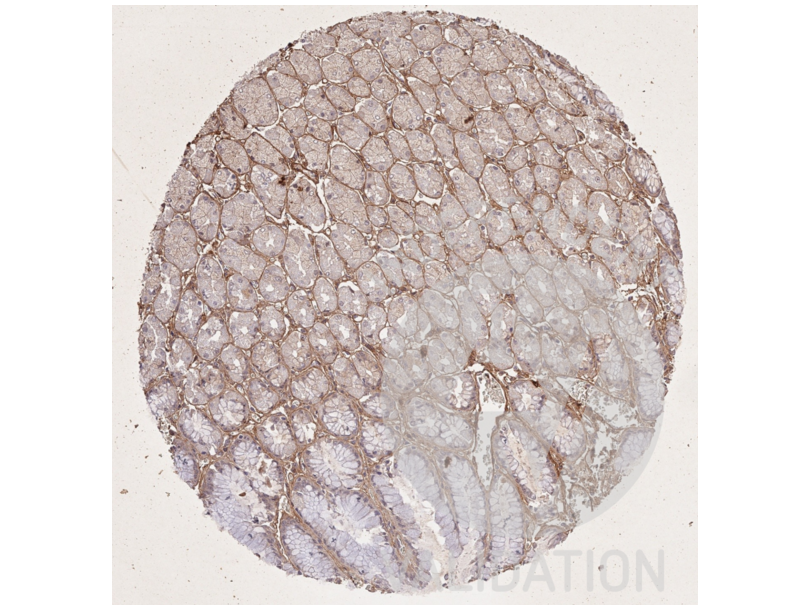
![IHC staining of the stomach mucosa with collagen type I antibody ABIN5596819 diluted 1:15 shows staining of basement membranes and the stomach mucosa.]() IHC staining of the stomach mucosa with collagen type I antibody ABIN5596819 diluted 1:15 shows staining of basement membranes and the stomach mucosa.
IHC staining of the stomach mucosa with collagen type I antibody ABIN5596819 diluted 1:15 shows staining of basement membranes and the stomach mucosa.
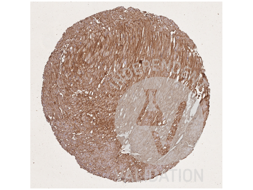
![IHC staining of stomach muscular wall with collagen type I antibody ABIN5596819 diluted 1:15 shows collagen I fibres surrounding smooth muscle cells.]() IHC staining of stomach muscular wall with collagen type I antibody ABIN5596819 diluted 1:15 shows collagen I fibres surrounding smooth muscle cells.
IHC staining of stomach muscular wall with collagen type I antibody ABIN5596819 diluted 1:15 shows collagen I fibres surrounding smooth muscle cells.
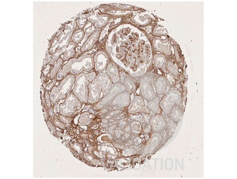
![IHC staining of kidney with collagen type I antibody ABIN5596819 diluted 1:15 shows intense staining of fibres surrounding tubuli and around blood vessels.]() IHC staining of kidney with collagen type I antibody ABIN5596819 diluted 1:15 shows intense staining of fibres surrounding tubuli and around blood vessels.
IHC staining of kidney with collagen type I antibody ABIN5596819 diluted 1:15 shows intense staining of fibres surrounding tubuli and around blood vessels.
![IHC staining of colorectal carcinoma with collagen type I antibody ABIN5596819 diluted 1:15 shows dense fibrillar collagen I deposits in the stroma. Cancer cells are collagen I negative.]() IHC staining of colorectal carcinoma with collagen type I antibody ABIN5596819 diluted 1:15 shows dense fibrillar collagen I deposits in the stroma. Cancer cells are collagen I negative.
Protokoll
IHC staining of colorectal carcinoma with collagen type I antibody ABIN5596819 diluted 1:15 shows dense fibrillar collagen I deposits in the stroma. Cancer cells are collagen I negative.
Protokoll -
- Format
- Liquid
- Konzentration
- 1.0 mg/mL
- Buffer
-
Buffer: 0.02 M Potassium Phosphate, 0.15 M Sodium Chloride, pH 7.2
Stabilizer: None
Preservative: 0.01 % (w/v) Sodium Azide - Konservierungsmittel
- Sodium azide
- Vorsichtsmaßnahmen
- This product contains Sodium azide: a POISONOUS AND HAZARDOUS SUBSTANCE which should be handled by trained staff only.
- Lagerung
- 4 °C,-20 °C
- Informationen zur Lagerung
- Store vial at 4° C prior to opening. This product is stable at 4° C as an undiluted liquid. Dilute only prior to immediate use. For extended storage, mix with an equal volume of glycerol, aliquot contents and freeze at -20° C or below. Avoid cycles of freezing and thawing.
- Haltbarkeit
- 12 months
-
-
: "A Scaffold-Free 3-D Co-Culture Mimics the Major Features of the Reverse Warburg Effect In Vitro." in: Cells, Vol. 9, Issue 8, (2020) (PubMed).
: "Attenuation of Flightless I Increases Human Pericyte Proliferation, Migration and Angiogenic Functions and Improves Healing in Murine Diabetic Wounds." in: International journal of molecular sciences, Vol. 21, Issue 16, (2020) (PubMed).
: "Parenchymal pericytes are not the major contributor of extracellular matrix in the fibrotic scar after stroke in male mice." in: Journal of neuroscience research, Vol. 98, Issue 5, pp. 826-842, (2020) (PubMed).
: "Effects of ASC Application on Endplate Regeneration Upon Glycerol-Induced Muscle Damage." in: Frontiers in molecular neuroscience, Vol. 13, pp. 107, (2020) (PubMed).
: "Effects of pirfenidone targeting the tumor microenvironment and tumor-stroma interaction as a novel treatment for non-small cell lung cancer." in: Scientific reports, Vol. 10, Issue 1, pp. 10900, (2020) (PubMed).
: "Recombinant spider silk protein eADF4(C16)-RGD coatings are suitable for cardiac tissue engineering." in: Scientific reports, Vol. 10, Issue 1, pp. 8789, (2020) (PubMed).
: "Asynchronous mixing of kidney progenitor cells potentiates nephrogenesis in organoids." in: Communications biology, Vol. 3, Issue 1, pp. 231, (2020) (PubMed).
: "Exogenous supply of Hsp47 triggers fibrillar collagen deposition in skin cell cultures in vitro." in: BMC molecular and cell biology, Vol. 21, Issue 1, pp. 22, (2020) (PubMed).
: "Lysophosphatidic Acid Induces ECM Production via Activation of the Mechanosensitive YAP/TAZ Transcriptional Pathway in Trabecular Meshwork Cells." in: Investigative ophthalmology & visual science, Vol. 59, Issue 5, pp. 1969-1984, (2019) (PubMed).
: "3D pulmospheres serve as a personalized and predictive multicellular model for assessment of antifibrotic drugs." in: JCI insight, Vol. 2, Issue 2, pp. e91377, (2019) (PubMed).
: "Lowering the culture temperature corrects collagen abnormalities caused by HSP47 gene knockout." in: Scientific reports, Vol. 9, Issue 1, pp. 17433, (2019) (PubMed).
: "Transcriptome analysis reveals autophagy as regulator of TGFβ/Smad-induced fibrogenesis in trabecular meshwork cells." in: Scientific reports, Vol. 9, Issue 1, pp. 16092, (2019) (PubMed).
: "Prion protein modulates endothelial to mesenchyme-like transition in trabecular meshwork cells: Implications for primary open angle glaucoma." in: Scientific reports, Vol. 9, Issue 1, pp. 13090, (2019) (PubMed).
: "Retrospective investigation of the prognostic value of the β1 integrin expression in patients with head and neck squamous cell carcinoma receiving primary radio(chemo)therapy." in: PLoS ONE, Vol. 13, Issue 12, pp. e0209479, (2019) (PubMed).
: "Intrinsic Immunity Shapes Viral Resistance of Stem Cells." in: Cell, Vol. 172, Issue 3, pp. 423-438.e25, (2019) (PubMed).
: "CMG2/ANTXR2 regulates extracellular collagen VI which accumulates in hyaline fibromatosis syndrome." in: Nature communications, Vol. 8, pp. 15861, (2018) (PubMed).
: "mTORC1 phosphorylates LARP6 to stimulate type I collagen expression." in: Scientific reports, Vol. 7, pp. 41173, (2018) (PubMed).
: "Transcriptome-based repurposing of apigenin as a potential anti-fibrotic agent targeting hepatic stellate cells." in: Scientific reports, Vol. 7, pp. 42563, (2018) (PubMed).
: "Dissecting the role of myeloid and mesenchymal fibroblasts in age-dependent cardiac fibrosis." in: Basic research in cardiology, Vol. 112, Issue 4, pp. 34, (2018) (PubMed).
: "Luteolin-Mediated Inhibition of Hepatic Stellate Cell Activation via Suppression of the STAT3 Pathway." in: International journal of molecular sciences, Vol. 19, Issue 6, (2018) (PubMed).
-
: "A Scaffold-Free 3-D Co-Culture Mimics the Major Features of the Reverse Warburg Effect In Vitro." in: Cells, Vol. 9, Issue 8, (2020) (PubMed).
-
- Target
- COL1A2/COL1A1
- Andere Bezeichnung
- COL1A1/A2
- Hintergrund
- Background: Collagens are highly conserved throughout evolution and are characterized by an uninterrupted ''Glycine-X-Y'' triplet repeat that is a necessary part of the triple helical structure. For these reasons, it is often extremely difficult to generate antibodies with specificities to collagens. The development of 'type' specific antibodies is dependent on NON-DENATURED three-dimensional epitopes. Rockland extensively purifies collagens for immunization from human and bovine placenta and cartilage by limited pepsin digestion and selective salt precipitation. This preparation results in a native conformation of the protein. Antibodies are isolated from rabbit antiserum and are extensively cross-adsorbed by immunoaffinity purification to produce 'type' specific antibodies. Greatly diminished reactivity and selectivity of these antibodies will result if denaturing and reducing conditions are used for SDS-PAGE and immunoblotting. Ideal for investigators involved in Cell Biology, Signal Transduction and Stem Cell research.
- Gen-ID
- 1277
- NCBI Accession
- NP_000079
- UniProt
- P02452
-


