STAT3 Antikörper (C-Term)
 (1 reference)
 (1 Validierung)
-
- Target Alle STAT3 Antikörper anzeigen
- STAT3 (Signal Transducer and Activator of Transcription 3 (Acute-Phase Response Factor) (STAT3))
-
Bindungsspezifität
- C-Term
-
Reaktivität
- Human
-
Wirt
- Kaninchen
-
Klonalität
- Polyklonal
-
Konjugat
- Dieser STAT3 Antikörper ist unkonjugiert
-
Applikation
- Western Blotting (WB), Immunofluorescence (IF), Immunohistochemistry (Paraffin-embedded Sections) (IHC (p)), Immunocytochemistry (ICC)
- Kreuzreaktivität
- Human, Maus, Pflanzen, Ratte
- Produktmerkmale
-
Rabbit Polyclonal antibody to STAT3 (signal transducer and activator of transcription 3 (acute-phase response factor))
STAT3 antibody [C3], C-term - Aufreinigung
- Purified by antigen-affinity chromatography.
- Güteklasse
- KO Validated
- Immunogen
- Carrier-protein conjugated synthetic peptide encompassing a sequence within the C-terminus region of human STAT3. The exact sequence is proprietary.
- Isotyp
- IgG
-
anti-Signal Transducer and Activator of Transcription 3 (Acute-Phase Response Factor) (STAT3) (C-Term) antibody
STAT3 Reaktivität: Human, Maus, Ratte WB, IHC, ELISA, IF, ICC Wirt: Kaninchen Polyclonal unconjugated
anti-Signal Transducer and Activator of Transcription 3 (Acute-Phase Response Factor) (STAT3) (pSer727) antibodySTAT3 Reaktivität: Human, Maus, Ratte WB, IHC, ELISA, IP, IF, ICC Wirt: Kaninchen Polyclonal unconjugated
anti-Signal Transducer and Activator of Transcription 3 (Acute-Phase Response Factor) (STAT3) (pTyr705) antibodySTAT3 Reaktivität: Human, Maus, Ratte WB, IHC, ELISA, IP, IF, ICC Wirt: Kaninchen Polyclonal unconjugated
anti-Signal Transducer and Activator of Transcription 3 (Acute-Phase Response Factor) (STAT3) (AA 640-770) antibodySTAT3 Reaktivität: Human WB, IHC, IF Wirt: Kaninchen Polyclonal unconjugated
anti-Signal Transducer and Activator of Transcription 3 (Acute-Phase Response Factor) (STAT3) (AA 233-330) antibodySTAT3 Reaktivität: Human, Maus, Ratte, Kaninchen WB, ELISA, IHC (p), FACS, IF (cc), IF (p), IHC (fro) Wirt: Kaninchen Polyclonal unconjugated
anti-Signal Transducer and Activator of Transcription 3 (Acute-Phase Response Factor) (STAT3) (pSer727) antibodySTAT3 Reaktivität: Human, Maus, Ratte WB, IHC (p), FACS, IF (cc) Wirt: Kaninchen Monoclonal 4G1 unconjugated
anti-Signal Transducer and Activator of Transcription 3 (Acute-Phase Response Factor) (STAT3) (AA 670-769) antibodySTAT3 Reaktivität: Human WB, ELISA, IF, PLA Wirt: Maus Monoclonal 4D6 unconjugated
anti-Signal Transducer and Activator of Transcription 3 (Acute-Phase Response Factor) (STAT3) (C-Term) antibodyVerified STAT3 Reaktivität: Human, Maus IHC, ELISA, IF, FACS Wirt: Ziege Polyclonal unconjugated
anti-Signal Transducer and Activator of Transcription 3 (Acute-Phase Response Factor) (STAT3) (pSer727) antibodySTAT3 Reaktivität: Human, Maus, Ratte, Affe WB, ELISA, IP, IF, IHC (p) Wirt: Kaninchen Polyclonal unconjugated
anti-Signal Transducer and Activator of Transcription 3 (Acute-Phase Response Factor) (STAT3) (pTyr705) antibodySTAT3 Reaktivität: Human, Maus, Ratte, Kaninchen, Schwein, Huhn WB, ELISA, IHC (p), FACS, IF (cc), IF (p), IHC (fro) Wirt: Kaninchen Polyclonal unconjugated
-
- Applikationshinweise
- WB: 1:500-1:3000. ICC/IF: 1:100-1:1000. Optimal dilutions/concentrations should be determined by the researcher. Not tested in other applications.
- Kommentare
-
Positive Control: Human ESC , OC3
Validation: Comparison, KO/KD, Orthogonal
- Beschränkungen
- Nur für Forschungszwecke einsetzbar
-
- by
- Gianluca Zambanini, Anna Nordin and Claudio Cantù; Cantù Lab, Gene Regulation during Development and Disease, Linköping University
- No.
- #104383
- Datum
- 07.12.2022
- Antigen
- STAT3
- Chargennummer
- 43873
- Validierte Anwendung
- Cleavage Under Targets and Release Using Nuclease
- Positivkontrolle
Polyclonal rabbit anti-H3K4me (antibodies-online, ABIN3023251)
- Negativkontrolle
Polyclonal guinea pig anti-rabbit IgG (antibodies-online, ABIN101961)
- Bewertung
Passed. ABIN2855865 allows for CUT&RUN targeted profiling of STAT3 in mouse forelimb tissues.
- Primärantikörper
- ABIN2855865
- Sekundärantikörper
- Full Protocol
- Cell harvest and nuclear extraction
- Dissect 3 Fore limbs (11.5 DAC) from mouse strain RjOrl:SWISS for each sample.
- Dissociate the tissue into single cells in TrypLE (Thermo Fisher Scientific) for 15 min at 37 °C.
- Centrifuge cell solution 5 min at 800 x g at RT.
- Remove the liquid carefully.
- Gently resuspend cells in 1 mL of Nuclear Extraction Buffer (20 mM HEPES-KOH pH 8.2, 20% Glycerol, 0.05% IGEPAL, 0.5 mM Spermidine, 10 mM KCl, Roche Complete Protease Inhibitor EDTA-free).
- Move the solution to a 2 mL centrifuge tube.
- Pellet the nuclei 800 x g for 5 min.
- Repeat the NE wash twice for a total of three washes.
- Resuspend the nuclei in 20 µL NE Buffer per sample.
- Concanavalin A beads preparation
- Prepare one 2 mL microcentrifuge tube.
- Gently resuspend the magnetic Concanavalin A Beads (antibodies-online, ABIN6952467).
- Pipette 20 µL Con A Beads slurry for each sample into the 2 mL microcentrifuge tube.
- Place the tube on a magnet stand until the fluid is clear. Remove the liquid carefully.
- Remove the microcentrifuge tube from the magnetic stand.
- Pipette 1 mL Binding Buffer (20 mM HEPES pH 7.5, 10 mM KCl, 1 mM CaCl2, 1 mM MnCl2) into the tube and resuspend ConA beads by gentle pipetting.
- Spin down the liquid from the lid with a quick pulse in a table-top centrifuge.
- Place the tubes on a magnet stand until the fluid is clear. Remove the liquid carefully.
- Remove the microcentrifuge tube from the magnetic stand.
- Repeat the wash twice for a total of three washes.
- Gently resuspend the ConA Beads in a volume of Binding Buffer corresponding to the original volume of bead slurry, i.e. 20 µL per sample.
- Nuclei immobilization – binding to Concanavalin A beads
- Carefully vortex the nuclei suspension and add 20 µL of the Con A beads in Binding Buffer to the cell suspension for each sample.
- Close tube tightly incubates 10 min at 4 °C.
- Put the 2 mL tube on the magnet stand and when the liquid is clear remove the supernatant.
- Resuspend the beads in 1 mL of EDTA Wash buffer (20 mM HEPES pH 7.5, 150 mM NaCl, 0.5 mM Spermidine, Roche Complete Protease Inhibitor EDTA-free, 2mM EDTA).
- Incubate 5 min at RT.
- Place the tube on the magnet stand and when the liquid is clear remove the supernatant.
- Resuspend the beads in 200 µl of Wash Buffer (20 mM HEPES pH 7.5, 150 mM NaCl, 0.5 mM Spermidine, Roche Complete Protease Inhibitor EDTA-free) per sample.
- Primary antibody binding
- Divide nuclei suspension into separate 200 µL PCR tubes, one for each antibody (150,000 cells per sample).
- Add 2 µL antibody (anti-STAT3 antibody ABIN2855865, anti-H3K4me positive control antibody ABIN3023251, and guinea pig anti-rabbit IgG negative control antibody ABIN101961) to the respective tube, corresponding to a 1:100 dilution.
- Incubate at 4 °C ON.
- Place the tubes on a magnet stand until the fluid is clear. Remove the liquid carefully.
- Remove the microcentrifuge tubes from the magnetic stand.
- Wash with 200 µL of Wash Buffer using a multichannel pipette to accelerate the process.
- Repeat the wash five times for a total of six washes.
- pAG-MNase Binding
- Prepare a 1.5 mL microcentrifuge tube containing 100 µL of pAG mix per sample (100 µL of wash buffer + 58.5 µg pAG-MNase per sample).
- Place the PCR tubes with the sample on a magnet stand until the fluid is clear. Remove the liquid carefully.
- Remove tubes from the magnetic stand.
- Resuspend the beads in 100 µL of pAG-MNase premix.
- Incubate 30 min at 4 °C.
- Place the tubes on a magnet stand until the fluid is clear. Remove the liquid carefully.
- Remove the microcentrifuge tubes from the magnetic stand.
- Wash with 200 µL of Wash Buffer using a multichannel pipette to accelerate the process.
- Repeat the wash five times for a total of six washes.
- Resuspend in 100 µL of Wash Buffer.
- MNase digestion and release of pAG-MNase-antibody-chromatin complexes
- Place PCR tubes on ice and allow to chill.
- Prepare a 1.5 mL microcentrifuge tube with 102 µl of 2 mM CaCl2 mix per sample (100 µl Wash Buffer + 2 µL 100 mM CaCl2) and let it chill on ice.
- Always in ice, place the samples on the magnetic rack and when the liquid is clear remove the supernatant.
- Resuspend the samples in 100 µl of the 2 mM CaCl2 mix and incubate in ice for exactly 30 min.
- Place the sample on the magnet stand and when the liquid is clear remove the supernatant.
- Resuspend the sample in 50 µl of 1x Urea STOP Buffer (8.5 M Urea, 100 mM NaCl, 2 mM EGTA, 2 mM EDTA, 0.5% IGEPAL).
- Incubate the samples 1h at 4°C.
- Transfer the supernatant containing the pAG-MNase-bound digested chromatin fragments to fresh 200 µl PCR tubes.
- DNA Clean up
- Take the Mag-Bind® TotalPure NGS beads (Omega Bio-Tek, M1378-01) from the storage and wait until they are at RT.
- Add 2x volume of beads to each sample (e.g. 100 µL of beads for 50 µL of sample).
- Incubate the beads and the sample for 15 min at RT.
- During incubation prepare fresh EtOH 80%.
- Place the PCR tubes on a magnet stand and when the liquid is clear remove the supernatant.
- Add 200 µl of fresh 80% EtOH to the sample without disturbing the beads (Important!!! Do NOT resuspend the beads or remove the tubes from the magnet stand or the sample will be lost).
- Incubate 30 sec at RT.
- Remove the EtOH from the sample.
- Repeat the wash with 80% EtOH.
- Resuspend the beads in 25 µL of 10 mM Tris-HCl pH 8.2.
- Incubate the sample for 2 min at RT.
- Repeat the 2x beads clean up as described before (this time with 50 µL of beads for each sample).
- Resuspend the beads + DNA in 20 µL of 10 mM Tris-HCl pH 8.2.
- Library preparation and sequencing
- Prepare Libraries using KAPA HyperPrep Kit using KAPA Dual-Indexed adapters according to protocol.
- Sequence samples on an Illumina NextSeq 500 sequencer, using a NextSeq 500/550 High Output Kit v2.5 (75 Cycles), 36 bp PE.
- Peak calling
- Trim reads using using bbTools bbduk (BBMap - Bushnell B. - sourceforge.net/projects/bbmap/) to remove adapters, artifacts and repeat sequences.
- Map aligned reads to the hg38 human genome using bowtie with options -m 1 -v 0 -I 0 -X 500.
- Use SAMtools to convert SAM files to BAM files and remove duplicates.
- Use BEDtools genomecov to produce Bedgraph files.
- Call peaks using SEACR with a 0.001 threshold and the option norm stringent.
- Anmerkungen
The protocol is published in Zambanini, G. et al. A New CUT&RUN Low Volume-Urea (LoV-U) protocol uncovers Wnt/β-catenin tissue-specific genomic targets. Development (2022). PMID 36355069
Validierung #104383 (Cleavage Under Targets and Release Using Nuclease)
 Validierungsbilder
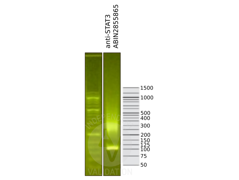
![Library profiles comparing fragment size distributions on an E-Gel EX 2% agarose gel (Thermo Fisher). Fragments obtained from CUT&RUN using anti-STAT3 antibody ABIN2855865 (right) after library preparation, compared to the E-Gel Sizing DNA Ladder (Thermo Fisher) (left).]() Library profiles comparing fragment size distributions on an E-Gel EX 2% agarose gel (Thermo Fisher). Fragments obtained from CUT&RUN using anti-STAT3 antibody ABIN2855865 (right) after library preparation, compared to the E-Gel Sizing DNA Ladder (Thermo Fisher) (left).
Library profiles comparing fragment size distributions on an E-Gel EX 2% agarose gel (Thermo Fisher). Fragments obtained from CUT&RUN using anti-STAT3 antibody ABIN2855865 (right) after library preparation, compared to the E-Gel Sizing DNA Ladder (Thermo Fisher) (left).
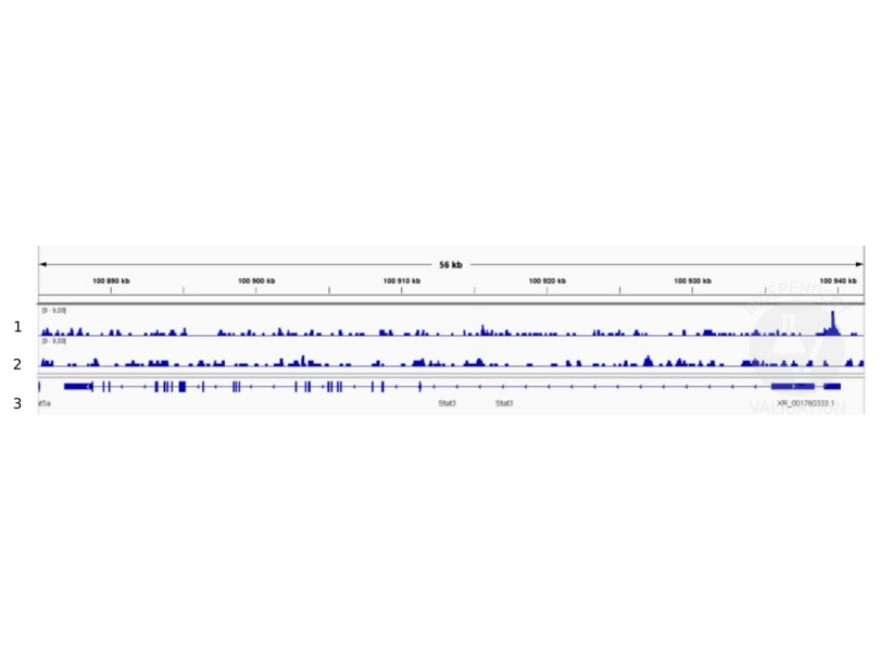
![1. Alignment tracks from CUT&RUN targeting STAT3 in mouse fore limb (11.5) cells using ABIN2855865, showing the STAT3 locus. 2. Alignment tracks using negative control IgG, ABIN1019613. 3. RefSeq Genes.]() 1. Alignment tracks from CUT&RUN targeting STAT3 in mouse fore limb (11.5) cells using ABIN2855865, showing the STAT3 locus. 2. Alignment tracks using negative control IgG, ABIN1019613. 3. RefSeq Genes.
Protokoll
1. Alignment tracks from CUT&RUN targeting STAT3 in mouse fore limb (11.5) cells using ABIN2855865, showing the STAT3 locus. 2. Alignment tracks using negative control IgG, ABIN1019613. 3. RefSeq Genes.
Protokoll -
- Format
- Liquid
- Konzentration
- 0.48 mg/mL
- Buffer
- 1XPBS ( pH 7), 1 % BSA, 20 % Glycerol, 0.025 % ProClin 300
- Konservierungsmittel
- ProClin
- Vorsichtsmaßnahmen
- This product contains ProClin: a POISONOUS AND HAZARDOUS SUBSTANCE which should be handled by trained staff only.
- Lagerung
- 4 °C,-20 °C
- Informationen zur Lagerung
- Store as concentrated solution. Centrifuge briefly prior to opening vial. For short-term storage (1-2 weeks), store at 4°C. For long-term storage, aliquot and store at -20°C or below. Avoid multiple freeze-thaw cycles.
-
-
: "ERK Activation Modulates Cancer Stemness and Motility of a Novel Mouse Oral Squamous Cell Carcinoma Cell Line." in: Cancers, Vol. 12, Issue 1, (2019) (PubMed).
-
: "ERK Activation Modulates Cancer Stemness and Motility of a Novel Mouse Oral Squamous Cell Carcinoma Cell Line." in: Cancers, Vol. 12, Issue 1, (2019) (PubMed).
-
- Target
- STAT3 (Signal Transducer and Activator of Transcription 3 (Acute-Phase Response Factor) (STAT3))
- Andere Bezeichnung
- signal transducer and activator of transcription 3 (STAT3 Produkte)
- Synonyme
- 1110034C02Rik antikoerper, AW109958 antikoerper, Aprf antikoerper, APRF antikoerper, HIES antikoerper, Xstat3 antikoerper, aprf antikoerper, hies antikoerper, stat3 antikoerper, wu:fc15d02 antikoerper, wu:fl59g06 antikoerper, z-Stat3 antikoerper, signal transducer and activator of transcription 3 antikoerper, signal transduction and activation of transcription 3 antikoerper, signal transducer and activator of transcription 3, gene 1 L homeolog antikoerper, signal transducer and activator of transcription 3 (acute-phase response factor) antikoerper, STAT3 antikoerper, stat3 antikoerper, Stat3 antikoerper, stat3.1.L antikoerper
- Hintergrund
-
The protein encoded by this gene is a member of the STAT protein family. In response to cytokines and growth factors, STAT family members are phosphorylated by the receptor associated kinases, and then form homo- or heterodimers that translocate to the cell nucleus where they act as transcription activators. This protein is activated through phosphorylation in response to various cytokines and growth factors including IFNs, EGF, IL5, IL6, HGF, LIF and BMP2. This protein mediates the expression of a variety of genes in response to cell stimuli, and thus plays a key role in many cellular processes such as cell growth and apoptosis. The small GTPase Rac1 has been shown to bind and regulate the activity of this protein. PIAS3 protein is a specific inhibitor of this protein. Three alternatively spliced transcript variants encoding distinct isoforms have been described.
Cellular Localization: Cytoplasm , Nucleus - Molekulargewicht
- 88 kDa
- Gen-ID
- 6774
- UniProt
- P40763
- Pathways
- JAK-STAT Signalweg, RTK Signalweg, Interferon-gamma Pathway, Neurotrophin Signalübertragung, Dopaminergic Neurogenesis, Response to Growth Hormone Stimulus, Carbohydrate Homeostasis, Stem Cell Maintenance, Hepatitis C, Protein targeting to Nucleus, Feeding Behaviour, CXCR4-mediated Signaling Events, Signaling of Hepatocyte Growth Factor Receptor
-


