HDAC1 Antikörper
 (7 Referenzen)
 (1 Validierung)
-
- Target Alle HDAC1 Antikörper anzeigen
- HDAC1 (Histone Deacetylase 1 (HDAC1))
-
Reaktivität
- Human
-
Wirt
- Kaninchen
-
Klonalität
- Polyklonal
-
Konjugat
- Dieser HDAC1 Antikörper ist unkonjugiert
-
Applikation
- Western Blotting (WB), Immunofluorescence (IF), Immunohistochemistry (Paraffin-embedded Sections) (IHC (p)), Immunoprecipitation (IP), Immunocytochemistry (ICC), Chromatin Immunoprecipitation (ChIP), Immunohistochemistry (Frozen Sections) (IHC (fro)), Immunohistochemistry (Whole Mount) (IHC (wm))
- Kreuzreaktivität
- Human, Maus, Ratte, Zebrafisch (Danio rerio)
- Produktmerkmale
-
Rabbit Polyclonal antibody to HDAC1 (histone deacetylase 1)
HDAC1 antibody - Aufreinigung
- Purified by antigen-affinity chromatography.
- Güteklasse
- KO Validated
- Immunogen
- Recombinant protein encompassing a sequence within the center region of human HDAC1. The exact sequence is proprietary.
- Isotyp
- IgG
-
anti-Histone Deacetylase 1 (HDAC1) (AA 466-482) antibody
HDAC1 Reaktivität: Human WB, ELISA, IHC Wirt: Kaninchen Polyclonal unconjugated
anti-Histone Deacetylase 1 (HDAC1) (AA 1-482) antibodyHDAC1 Reaktivität: Human WB, ELISA, IF, IHC (p), PLA Wirt: Maus Monoclonal 5C11 unconjugated
anti-Histone Deacetylase 1 (HDAC1) (AA 393-482) antibodyHDAC1 Reaktivität: Human WB, IHC, IF, IP Wirt: Kaninchen Polyclonal unconjugated
anti-Histone Deacetylase 1 (HDAC1) (AA 1-482) antibodyHDAC1 Reaktivität: Human WB, IHC, IP, ICC Wirt: Maus Monoclonal E22 unconjugated
anti-Histone Deacetylase 1 (HDAC1) (AA 1-482) antibodyHDAC1 Reaktivität: Human ELISA, IHC, IF, ChIP Wirt: Kaninchen Polyclonal unconjugated
anti-Histone Deacetylase 1 (HDAC1) (AA 1-482) antibodyHDAC1 Reaktivität: Human WB, ELISA, IF, IHC (p) Wirt: Maus Monoclonal 5A11 unconjugated
anti-Histone Deacetylase 1 (HDAC1) antibodyKO Validated HDAC1 Reaktivität: Human WB, IHC, IF Wirt: Kaninchen Monoclonal unconjugated
anti-Histone Deacetylase 1 (HDAC1) (C-Term) antibodyHDAC1 Reaktivität: Human, Maus WB, ELISA, IF, ChIP, ChIP-seq Wirt: Kaninchen Polyclonal unconjugated
anti-Histone Deacetylase 1 (HDAC1) (AA 207-412) antibodyHDAC1 Reaktivität: Human WB, ELISA, IHC, IF Wirt: Kaninchen Polyclonal unconjugated
anti-Histone Deacetylase 1 (HDAC1) (AA 449-482), (C-Term) antibodyHDAC1 Reaktivität: Human, Maus WB, IF Wirt: Kaninchen Polyclonal RB5642 unconjugated
-
- Applikationshinweise
- WB: 1:500-1:3000. ICC/IF: 1:100-1:1000. IHC-P: 1:100-1:1000. IP: 1:100-1:500. Optimal dilutions/concentrations should be determined by the researcher. Not tested in other applications.
- Kommentare
-
Positive Control: 293T , A431 , HeLa , HepG2 , U87-MG , SK-N-SH , Rat-2 , NIH3T3 , DDDDK-tagged HDAC1-transfected 293T
Validation: KO/KD, Orthogonal, Overexpression
- Beschränkungen
- Nur für Forschungszwecke einsetzbar
-
- by
- Gianluca Zambanini, Anna Nordin and Claudio Cantù; Cantù Lab, Gene Regulation during Development and Disease, Linköping University
- No.
- #104404
- Datum
- 28.02.2022
- Antigen
- HDAC1
- Chargennummer
- Validierte Anwendung
- Cleavage Under Targets and Release Using Nuclease
- Positivkontrolle
Polyclonal rabbit anti-H3K4me (antibodies-online, ABIN3023251)
- Negativkontrolle
Polyclonal guinea pig anti-rabbit IgG (antibodies-online, ABIN101961)
- Bewertung
Passed. ABIN2854776 allows for HDAC1 targeted digestion using CUT&RUN in mouse fore limbs (11.5) cells.
- Primärantikörper
- ABIN2854776
- Sekundärantikörper
- Full Protocol
- Cell harvest and nuclear extraction
- Dissect 3 Fore limbs (11.5 DAC) from mouse strain RjOrl:SWISS for each sample.
- Dissociate the tissue into single cells in TrypLE for 15 min at 37 °C.
- Centrifuge cell solution 5 min at 800 x g at RT.
- Remove the liquid carefully.
- Gently resuspend cells in 1 mL of Nuclear Extraction Buffer (20 mM HEPES-KOH pH 8.2, 20% Glycerol, 0,05% IGEPAL, 0.5 mM Spermidine, 10 mM KCl, Roche Complete Protease Inhibitor EDTA-free).
- Move the solution to a 2 mL centrifuge tube.
- Pellet the nuclei 800 x g for 5 min.
- Repeat the NE wash twice for a total of three washes.
- Resuspend the nuclei in 20 µL NE Buffer per sample.
- Concanavalin A beads preparation
- Prepare one 2 mL microcentrifuge tube.
- Gently resuspend the magnetic Concanavalin A Beads (antibodies-online, ABIN6952467).
- Pipette 20 µL Con A Beads slurry for each sample into the 2 mL microcentrifuge tube.
- Place the tube on a magnet stand until the fluid is clear. Remove the liquid carefully.
- Remove the microcentrifuge tube from the magnetic stand.
- Pipette 1 mL Binding Buffer (20 mM HEPES pH 7.5, 10 mM KCl, 1 mM CaCl2, 1 mM MnCl2) into the tube and resuspend ConA beads by gentle pipetting.
- Spin down the liquid from the lid with a quick pulse in a table-top centrifuge.
- Place the tubes on a magnet stand until the fluid is clear. Remove the liquid carefully.
- Remove the microcentrifuge tube from the magnetic stand.
- Repeat the wash twice for a total of three washes.
- Gently resuspend the ConA Beads in a volume of Binding Buffer corresponding to the original volume of bead slurry, i.e. 20 µL per sample.
- Nuclei immobilization – binding to Concanavalin A beads
- Carefully vortex the nuclei suspension and add 20 µL of the Con A beads in Binding Buffer to the cell suspension for each sample.
- Close tube tightly incubates 10 min at 4 °C.
- Put the 2 mL tube on the magnet stand and when the liquid is clear remove the supernatant.
- Resuspend the beads in 1 mL of EDTA Wash buffer (20 mM HEPES pH 7.5, 150 mM NaCl, 0.5 mM Spermidine, Roche Complete Protease Inhibitor EDTA-free, 2mM EDTA).
- Incubate 5 min at RT.
- Place the tube on the magnet stand and when the liquid is clear remove the supernatant.
- Resuspend the beads in 200 µl of Wash Buffer (20 mM HEPES pH 7.5, 150 mM NaCl, 0.5 mM Spermidine, Roche Complete Protease Inhibitor EDTA-free) per sample.
- Primary antibody binding
- Divide nuclei suspension into separate 200 µL PCR tubes, one for each antibody.
- Add 2 µL antibody (anti-HDAC1 antibody ABIN2854776, anti-H3K27me3 antibody positive control ABIN6923144, and guinea pig anti-rabbit IgG negative control antibody ABIN101961) to the respective tube, corresponding to a 1:100 dilution.
- Incubate at 4 °C ON.
- Place the tubes on a magnet stand until the fluid is clear. Remove the liquid carefully.
- Remove the microcentrifuge tubes from the magnetic stand.
- Wash with 200 µL of Wash Buffer using a multichannel pipette to accelerate the process.
- Repeat the wash five times for a total of six washes.
- pAG-MNase Binding
- Prepare a 1.5 mL microcentrifuge tube containing 100 µL of pAG mix per sample (100 µL of wash buffer + 58.5 µg pAG-MNase per sample).
- Place the PCR tubes with the sample on a magnet stand until the fluid is clear. Remove the liquid carefully.
- Remove tubes from the magnetic stand.
- Resuspend the beads in 100 µL of pAG-MNase premix.
- Incubate 30 min at 4 °C.
- Place the tubes on a magnet stand until the fluid is clear. Remove the liquid carefully.
- Remove the microcentrifuge tubes from the magnetic stand.
- Wash with 200 µL of Wash Buffer using a multichannel pipette to accelerate the process.
- Repeat the wash five times for a total of six washes.
- Resuspend in 100 µL of Wash Buffer.
- MNase digestion and release of pAG-MNase-antibody-chromatin complexes
- Place PCR tubes on ice and allow to chill.
- Prepare a 1.5 mL microcentrifuge tube with 102 µl of 2 mM CaCl2 mix per sample (100 µl Wash Buffer + 2 µL 100 mM CaCl2) and let it chill on ice.
- Always in ice, place the samples on the magnetic rack and when the liquid is clear remove the supernatant.
- Resuspend the samples in 100 µl of the 2 mM CaCl2 mix and incubate in ice for exactly 30 min.
- Place the sample on the magnet stand and when the liquid is clear remove the supernatant.
- Resuspend the sample in 50 µl of 1x Urea STOP Buffer (8.5 M Urea, 100 mM NaCl, 2 mM EGTA, 2 mM EDTA, 0,5% IGEPAL).
- Incubate the samples 1h at 4°C.
- Transfer the supernatant containing the pAG-MNase-bound digested chromatin fragments to fresh 200 µl PCR tubes.
- DNA Clean up
- Take the Mag-Bind® TotalPure NGS beads (Omega Bio-Tek, M1378-01) from the storage and wait until they are at RT.
- Add 2x volume of beads to each sample (e.g. 100 µL of beads for 50 µL of sample).
- Incubate the beads and the sample for 15 min at RT.
- During incubation prepare fresh EtOH 80%.
- Place the PCR tubes on a magnet stand and when the liquid is clear remove the supernatant.
- Add 200 µl of fresh 80% EtOH to the sample without disturbing the beads (Important!!! Do NOT resuspend the beads or remove the tubes from the magnet stand or the sample will be lost).
- Incubate 30 sec at RT.
- Remove the EtOH from the sample.
- Repeat the wash with 80% EtOH.
- Resuspend the beads in 25 µL of 10 mM Tris.
- Incubate the sample for 2 min at RT.
- Repeat the 2x beads clean up as described before (this time with 50 µL of beads for each sample).
- Resuspend the beads + DNA in 20 µL of 10 mM Tris.
- Library preparation and sequencing
- Prepare Libraries using KAPA HyperPrep Kit using KAPA Dual-Indexed adapters according to protocol.
- Sequence samples on an Illumina NextSeq 500 sequencer, using a NextSeq 500/550 High Output Kit v2.5 (75 Cycles), 36 bp PE.
- Peak calling
- Trim reads using using bbTools bbduk (BBMap - Bushnell B. - sourceforge.net/projects/bbmap/) to remove adapters, artifacts and repeat sequences.
- Map aligned reads to the hg38 human genome using bowtie with options -m 1 -v 0 -I 0 -X 500.
- Use SAMtools to convert SAM files to BAM files and remove duplicates.
- Use BEDtools genomecov to produce Bedgraph files.
- Call peaks using SEACR with a 0.001 threshold and the option norm stringent.
- Anmerkungen
The protocol is published in Zambanini, G. et al. A New CUT&RUN Low Volume-Urea (LoV-U) protocol uncovers Wnt/β-catenin tissue-specific genomic targets. bioRxiv (2022). https://doi.org/10.1101/2022.07.06.498999
Validierung #104404 (Cleavage Under Targets and Release Using Nuclease)
 Validierungsbilder
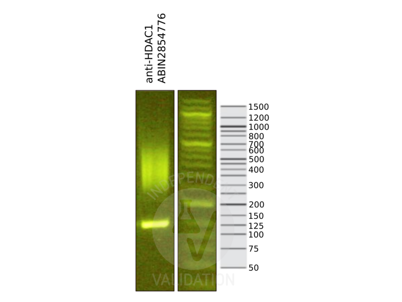
![Library profiles comparing fragment size distributions on an E-Gel EX 2% agarose gel (Thermo Fisher). Fragments obtained from CUT&RUN using anti-HDAC1 antibody ABIN2854776 after library preparation, compared to the E-Gel Sizing DNA Ladder (Thermo Fisher).]() Library profiles comparing fragment size distributions on an E-Gel EX 2% agarose gel (Thermo Fisher). Fragments obtained from CUT&RUN using anti-HDAC1 antibody ABIN2854776 after library preparation, compared to the E-Gel Sizing DNA Ladder (Thermo Fisher).
Library profiles comparing fragment size distributions on an E-Gel EX 2% agarose gel (Thermo Fisher). Fragments obtained from CUT&RUN using anti-HDAC1 antibody ABIN2854776 after library preparation, compared to the E-Gel Sizing DNA Ladder (Thermo Fisher).
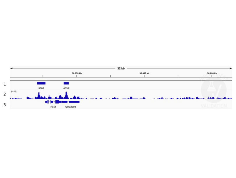
![1. Alignment tracks from CUT&RUN targeting HDAC1 in Mouse fore limb (11.5) cells using anti-HDAC1 antibody ABIN2854776. 2. Peaks called by SEACR from CUT&RUN data using anti HDAC1 ABIN2854776. 3. RefSeq Genes.]() 1. Alignment tracks from CUT&RUN targeting HDAC1 in Mouse fore limb (11.5) cells using anti-HDAC1 antibody ABIN2854776. 2. Peaks called by SEACR from CUT&RUN data using anti HDAC1 ABIN2854776. 3. RefSeq Genes.
Protokoll
1. Alignment tracks from CUT&RUN targeting HDAC1 in Mouse fore limb (11.5) cells using anti-HDAC1 antibody ABIN2854776. 2. Peaks called by SEACR from CUT&RUN data using anti HDAC1 ABIN2854776. 3. RefSeq Genes.
Protokoll -
- Format
- Liquid
- Konzentration
- 1 mg/mL
- Buffer
- 1XPBS ( pH 7), 20 % Glycerol, 0.01 % Thimerosal
- Konservierungsmittel
- Thimerosal (Merthiolate)
- Vorsichtsmaßnahmen
- This product contains Thimerosal (Merthiolate): a POISONOUS AND HAZARDOUS SUBSTANCE which should be handled by trained staff only.
- Lagerung
- 4 °C,-20 °C
- Informationen zur Lagerung
- Store as concentrated solution. Centrifuge briefly prior to opening vial. For short-term storage (1-2 weeks), store at 4°C. For long-term storage, aliquot and store at -20°C or below. Avoid multiple freeze-thaw cycles.
-
-
: "A new cut&run low volume-urea (LoV-U) protocol optimized for transcriptional co-factors uncovers Wnt/b-catenin tissue-specific genomic targets." in: Development (Cambridge, England), (2022) (PubMed).
: "Differential Expression of Multiple Disease-Related Protein Groups Induced by Valproic Acid in Human SH-SY5Y Neuroblastoma Cells." in: Brain sciences, Vol. 10, Issue 8, (2020) (PubMed).
: "Genome-wide kinetic properties of transcriptional bursting in mouse embryonic stem cells." in: Science advances, Vol. 6, Issue 25, pp. eaaz6699, (2020) (PubMed).
: "Nifedipine Exacerbates Lipogenesis in the Kidney via KIM-1, CD36, and SREBP Upregulation: Implications from an Animal Model for Human Study." in: International journal of molecular sciences, Vol. 21, Issue 12, (2020) (PubMed).
: "Overexpression of peptidase inhibitor 16 attenuates angiotensin II-induced cardiac fibrosis via regulating HDAC1 of cardiac fibroblasts." in: Journal of cellular and molecular medicine, Vol. 24, Issue 9, pp. 5249-5259, (2020) (PubMed).
: "The chromatin remodeler RSF1 controls centromeric histone modifications to coordinate chromosome segregation." in: Nature communications, Vol. 9, Issue 1, pp. 3848, (2019) (PubMed).
: "Inhibition of HDAC3- and HDAC6-Promoted Survivin Expression Plays an Important Role in SAHA-Induced Autophagy and Viability Reduction in Breast Cancer Cells." in: Frontiers in pharmacology, Vol. 7, pp. 81, (2016) (PubMed).
-
: "A new cut&run low volume-urea (LoV-U) protocol optimized for transcriptional co-factors uncovers Wnt/b-catenin tissue-specific genomic targets." in: Development (Cambridge, England), (2022) (PubMed).
-
- Target
- HDAC1 (Histone Deacetylase 1 (HDAC1))
- Andere Bezeichnung
- histone deacetylase 1 (HDAC1 Produkte)
- Synonyme
- GON-10 antikoerper, HD1 antikoerper, RPD3 antikoerper, RPD3L1 antikoerper, CG7471 antikoerper, DHDAC1 antikoerper, DRpd3 antikoerper, DmHDAC1 antikoerper, Dmel\\CG7471 antikoerper, E(var)3-64BC antikoerper, HDAC antikoerper, HDAC-1 antikoerper, HDAC1 antikoerper, Hdac1 antikoerper, Rpd3/HDAC antikoerper, Su(var)3-26 antikoerper, Su(var)326 antikoerper, Su(var)328 antikoerper, dHDAC-1 antikoerper, dHDAC1 antikoerper, dRPD3 antikoerper, dRpd3 antikoerper, dmHDA401 antikoerper, drpd3 antikoerper, hdac1 antikoerper, l(3)04556 antikoerper, l(3)64Cc antikoerper, rpd3 antikoerper, rpd[3] antikoerper, gon-10 antikoerper, hdac1b antikoerper, rpd3l1 antikoerper, Rpd3 antikoerper, GB14706 antikoerper, hdac1-b antikoerper, chunp6919 antikoerper, hdac-1 antikoerper, mp:zf637-2-001987 antikoerper, wu:fb19h11 antikoerper, wu:fi06f03 antikoerper, zgc:101582 antikoerper, zgc:65818 antikoerper, Hdac1-ps antikoerper, MommeD5 antikoerper, ARABIDOPSIS HISTONE DEACETYLASE 1 antikoerper, ARABIDOPSIS HISTONE DEACETYLASE 19 antikoerper, ATHD1 antikoerper, ATHDA19 antikoerper, ATRPD3A antikoerper, F20D10.250 antikoerper, F20D10_250 antikoerper, HDA1 antikoerper, HDA19 antikoerper, HISTONE DEACETYLASE antikoerper, HISTONE DEACETYLASE 19 antikoerper, HISTONE DEACETYLASE19 antikoerper, RPD3A antikoerper, histone deacetylase 1 antikoerper, HDM antikoerper, ab21 antikoerper, hdac1a antikoerper, histone deacetylase 1 antikoerper, Histone deacetylase 1 antikoerper, histone deacetylase antikoerper, histone deacetylase 1 S homeolog antikoerper, histone deacetylase Rpd3 antikoerper, histone deacetylase 1 L homeolog antikoerper, HDAC1 antikoerper, hdac1.S antikoerper, LOC411503 antikoerper, hdac1 antikoerper, Hdac1 antikoerper, HD1 antikoerper, hda-1 antikoerper, hdac1.L antikoerper, LOC748850 antikoerper
- Hintergrund
-
Histone acetylation and deacetylation, catalyzed by multisubunit complexes, play a key role in the regulation of eukaryotic gene expression. The protein encoded by this gene belongs to the histone deacetylase/acuc/apha family and is a component of the histone deacetylase complex. It also interacts with retinoblastoma tumor-suppressor protein and this complex is a key element in the control of cell proliferation and differentiation. Together with metastasis-associated protein-2, it deacetylates p53 and modulates its effect on cell growth and apoptosis.
Cellular Localization: Nucleus - Molekulargewicht
- 55 kDa
- Gen-ID
- 3065
- UniProt
- Q13547
- Pathways
- Neurotrophin Signalübertragung, Intracellular Steroid Hormone Receptor Signaling Pathway, Regulation of Intracellular Steroid Hormone Receptor Signaling, Mitotic G1-G1/S Phases, Regulation of Muscle Cell Differentiation, Skeletal Muscle Fiber Development, Negative Regulation of intrinsic apoptotic Signaling, Embryonic Body Morphogenesis
-


