SLC18A3 Antikörper (C-Term)
 (4 Referenzen)
 (1 Validierung)
-
- Target Alle SLC18A3 Antikörper anzeigen
- SLC18A3 (Solute Carrier Family 18 (Vesicular Acetylcholine), Member 3 (SLC18A3))
-
Bindungsspezifität
- AA 475-530, C-Term
-
Reaktivität
- Ratte, Maus
-
Wirt
-
Meerschweinchen
-
Klonalität
- Polyklonal
-
Konjugat
- Dieser SLC18A3 Antikörper ist unkonjugiert
-
Applikation
- Western Blotting (WB), Immunohistochemistry (IHC)
- Spezifität
- Specific for VAChT.
- Aufreinigung
- Affinity purified with the immunogen. Guinea pig serum albumin was added for stabilization.
- Immunogen
- Strep-Tag® fusion protein of the C-terminal part of rat VAChT (aa 475-530).
-
anti-Solute Carrier Family 18 (Vesicular Acetylcholine), Member 3 (SLC18A3) (AA 521-532) antibody
SLC18A3 Reaktivität: Human WB, IHC, IF, ICC Wirt: Maus Monoclonal S6-38 unconjugated
anti-Solute Carrier Family 18 (Vesicular Acetylcholine), Member 3 (SLC18A3) (AA 521-532) antibody (HRP)SLC18A3 Reaktivität: Human WB, IHC, IF, ICC Wirt: Maus Monoclonal S6-38 HRP
anti-Solute Carrier Family 18 (Vesicular Acetylcholine), Member 3 (SLC18A3) (AA 521-532) antibody (FITC)SLC18A3 Reaktivität: Human WB, IHC, IF, ICC Wirt: Maus Monoclonal S6-38 FITC
anti-Solute Carrier Family 18 (Vesicular Acetylcholine), Member 3 (SLC18A3) (AA 1-150) antibodySLC18A3 Reaktivität: Human WB, IHC, IF Wirt: Kaninchen Polyclonal unconjugated
anti-Solute Carrier Family 18 (Vesicular Acetylcholine), Member 3 (SLC18A3) (AA 521-532) antibody (Biotin)SLC18A3 Reaktivität: Human WB, IHC, IF, ICC Wirt: Maus Monoclonal S6-38 Biotin
anti-Solute Carrier Family 18 (Vesicular Acetylcholine), Member 3 (SLC18A3) (AA 521-532) antibody (PE)SLC18A3 Reaktivität: Human WB, IHC, IF, ICC Wirt: Maus Monoclonal S6-38 PE
anti-Solute Carrier Family 18 (Vesicular Acetylcholine), Member 3 (SLC18A3) (AA 521-532) antibody (APC)SLC18A3 Reaktivität: Human WB, IHC, IF, ICC Wirt: Maus Monoclonal S6-38 APC
anti-Solute Carrier Family 18 (Vesicular Acetylcholine), Member 3 (SLC18A3) (AA 521-532) antibody (PerCP)SLC18A3 Reaktivität: Human WB, IHC, IF, ICC Wirt: Maus Monoclonal S6-38 PerCP
anti-Solute Carrier Family 18 (Vesicular Acetylcholine), Member 3 (SLC18A3) (AA 521-532) antibody (Atto 488)SLC18A3 Reaktivität: Human WB, IHC, IF, ICC Wirt: Maus Monoclonal S6-38 Atto 488
anti-Solute Carrier Family 18 (Vesicular Acetylcholine), Member 3 (SLC18A3) (AA 521-532) antibody (Atto 594)SLC18A3 Reaktivität: Human WB, IHC, IF, ICC Wirt: Maus Monoclonal S6-38 Atto 594
-
- Applikationshinweise
-
WB: 1 : 500 up to 1 : 1000 (AP staining)
IP: not tested yet
ICC: not tested yet
IHC: 1 : 100 up to 1 : 300 - Kommentare
-
This antibody is less sensitive compared to the rabbit antibody. VAChT aggregates after boiling, making it necessary to run SDS-PAGE only with non-boiled samples.
- Beschränkungen
- Nur für Forschungszwecke einsetzbar
-
- by
- Martinelli Lab, Neuroscience Department, UConn Health
- No.
- #101568
- Datum
- 05.09.2017
- Antigen
- SLC18A3
- Chargennummer
- 139105/6
- Validierte Anwendung
- Immunofluorescence
- Positivkontrolle
- Organ of Corti tissue dissected from the cochlea of the inner ear, from a WT mouse, C57BL/6 strain, 10 weeks old
- Negativkontrolle
- Primary and secondary only antibody controls
- Bewertung
- Passed. ABIN1742304 specifically recognizes SLC18A3 with little background signal in murine organ of Corti tissue dissected from the cochlea of the inner ear.
- Primärantikörper
- ABIN1742304
- Sekundärantikörper
- goat anti-guinea pig AF633 conjugated antibody (Life Technologies, A21105, lot 1812311)
- Full Protocol
- Dissect organ of Corti from the mouse cochlea in the inner ear as described in Maison, Liberman, and Liberman (2016).
- Cryoprotect and freeze/thaw to permeabilized the tissue:
- Transfer cochlear pieces to a 5ml disposable cup with approximately 1ml of 30% sucrose in 100mM phosphate buffer (PB) at RT.
- Incubate tissue on a shaker for 15min at RT.
- Wash tisse with 30% sucrose in PB at RT.
- Incubate tissue on a shaker for 15min at RT.
- Place cup on dry ice untill contents freeze completely.
- Allow cup to thaw at RT.
- Pipet out the sucrose solution and wash tissue 3x for 15min with PBS containing 0.1% triton X-100 on a shaker at RT.
- Block tissue with 5% goat serum containing 0.3% Triton-X on a shaker for 30-60 min at RT.
- Pipet out PBS + detergent and add blocking solution (supplier, product no, lot no).
- Cut cap off a 1.5ml microcentrifuge tube and transfer pieces in the blocking solution to the flipped upside-down cap.
- Incubate tissue in the flipped upside-down caps with 100µl primary
- guinea pig anti-SLC18A3 antibody (antibodies-online, ABIN1742304, lot 139105/6) diluted 1:300
- chicken anti-Parvalbumin (Synaptic Systems, 195006) diluted 1:400
- in blocking solution ON at RT on an agitator. Fasten tubes onto the caps and and protect them from the light.
- Pipet out the primaries.
- Rinse tissue 3x for a total of 10 min with PBS containing 0.1 % Triton X-100.
- Incubate tissue in the flipped upside-down caps with 100µl secondary
- goat anti-guinea pig AF633 conjugated antibody (Life Technologies, A21105, lot 1812311)
- goat anti-chicken AF488 conjugated antibody (Life Technologies, A11039, lot 1812246)
- diluted 1:300 in blocking solution for 1h at RT away from the light.
- Rinse tissue 3x for a total of 10min with PBS.
- Transfer tissue pieces onto a slide with stereocilia facing up.
- Add mounting Fluoromount-G with DAPI mounting medium (ThermoFisher Scientific, 00-4959-52, lot B2215-N915) then coverslip.
- Image acquisition on a Zeiss Axiovert epifluorescence with with a 63x objective, using a Zeiss ApoTome for optical sectioning.
- Anmerkungen
The observed signal on outer hair cells in the organ of Corti for ABIN1742304 appears indistinguishable to the published and expected signal documented in numerous publications (see e.g. figure 4 in Maison, Liberman, and Liberman (2016)).
No signal was observed in either of the negative controls. ABIN1742304 worked very well at 1:300 dilution. It could probably be further diluted and still achieve acceptable signal to noise ratio.
No signal was observed with the secondary antibody only negative control.
Validierung #101568 (Immunofluorescence)
 Validierungsbilder
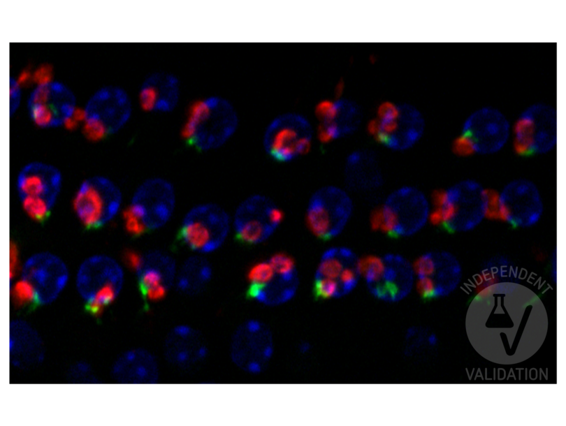
![In this photograph of the 3 rows of cochlear outer hair cells taken with 63x objective, nuclei are labeled with DAPI (blue), SLC18A3 is labeled with ABIN1742304 (red), and green marks the parvalbumin signal. Note that SLC18A3-positive synapses are adjacent to the parvalbumin-positive synapses, as expected since SLC18A3 marks efferent synapses and parvalbumin marks afferent synapses. These two synapses are known to be adjacent on outer hair cells.]() In this photograph of the 3 rows of cochlear outer hair cells taken with 63x objective, nuclei are labeled with DAPI (blue), SLC18A3 is labeled with ABIN1742304 (red), and green marks the parvalbumin signal. Note that SLC18A3-positive synapses are adjacent to the parvalbumin-positive synapses, as expected since SLC18A3 marks efferent synapses and parvalbumin marks afferent synapses. These two synapses are known to be adjacent on outer hair cells.
Protokoll
In this photograph of the 3 rows of cochlear outer hair cells taken with 63x objective, nuclei are labeled with DAPI (blue), SLC18A3 is labeled with ABIN1742304 (red), and green marks the parvalbumin signal. Note that SLC18A3-positive synapses are adjacent to the parvalbumin-positive synapses, as expected since SLC18A3 marks efferent synapses and parvalbumin marks afferent synapses. These two synapses are known to be adjacent on outer hair cells.
Protokoll -
- Format
- Lyophilized
- Rekonstitution
- For reconstitution add 50 µL H2O to get a 1mg/ml solution of antibody in PBS. Then aliquot and store at -20 °C until use.
- Buffer
- PBS
- Handhabung
- Affinity purified antibodies are less robust than antisera, since protease inhibitors are also removed during purification. Hence, storage at 4 °C for prolonged periods (i.e. several weeks), is not recommended.
- Lagerung
- -20 °C
- Informationen zur Lagerung
- Unlabeled lyophilized antibodies are stable in this form without loss of quality at ambient temperatures for several weeks or even months. They can be stored at 4°C for several years. Lyophilized antibodies must not be stored in the freezer, they may be destroyed!
-
-
: "Glutamatergic and central cholinergic dysfunction in the CA1, CA2 and CA3 fields on spatial learning and memory in chronic cerebral ischemia-Induced vascular dementia of rats." in: Neuroscience letters, Vol. 620, pp. 169-176, (2016) (PubMed).
: "Deletion of neurturin impairs development of cholinergic nerves and heart rate control in postnatal mouse hearts." in: Physiological reports, Vol. 4, Issue 9, (2016) (PubMed).
: "An animal model of Miller Fisher syndrome: Mitochondrial hydrogen peroxide is produced by the autoimmune attack of nerve terminals and activates Schwann cells." in: Neurobiology of disease, Vol. 96, pp. 95-104, (2016) (PubMed).
: "Neuregulin-1 is concentrated in the postsynaptic subsurface cistern of C-bouton inputs to α-motoneurons and altered during motoneuron diseases." in: FASEB journal : official publication of the Federation of American Societies for Experimental Biology, Vol. 28, Issue 8, pp. 3618-32, (2014) (PubMed).
-
: "Glutamatergic and central cholinergic dysfunction in the CA1, CA2 and CA3 fields on spatial learning and memory in chronic cerebral ischemia-Induced vascular dementia of rats." in: Neuroscience letters, Vol. 620, pp. 169-176, (2016) (PubMed).
-
- Target
- SLC18A3 (Solute Carrier Family 18 (Vesicular Acetylcholine), Member 3 (SLC18A3))
- Andere Bezeichnung
- VAChT (SLC18A3 Produkte)
- Synonyme
- VACht antikoerper, rVAT antikoerper, Slc18a3 antikoerper, MGC64220 antikoerper, VACHT antikoerper, VAChT antikoerper, VAT antikoerper, SLC18A3 antikoerper, CG12345 antikoerper, CG32848 antikoerper, CT41182 antikoerper, Dmel\\CG32848 antikoerper, Vacht antikoerper, vAChT antikoerper, vacht antikoerper, VAChT-A antikoerper, zgc:153442 antikoerper, solute carrier family 18 member A3 antikoerper, solute carrier family 18 (vesicular acetylcholine transporter), member 3b antikoerper, solute carrier family 18 (vesicular monoamine), member 3 antikoerper, solute carrier family 18 (vesicular acetylcholine transporter), member 3 antikoerper, Vesicular acetylcholine transporter antikoerper, solute carrier family 18 (vesicular acetylcholine transporter), member 3a antikoerper, Slc18a3 antikoerper, slc18a3b antikoerper, SLC18A3 antikoerper, slc18a3 antikoerper, VAChT antikoerper, slc18a3a antikoerper
-


